KCNA5
Potassium voltage-gated channel, shaker-related subfamily, member 5, also known as KCNA5 or Kv1.5, is a protein that in humans is encoded by the KCNA5 gene.[5]
Function
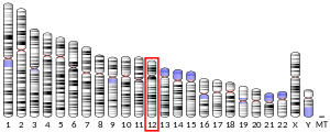
Potassium channels represent the most complex class of voltage-gated ion channels from both functional and structural standpoints. KCNA5 encodes a member of the potassium channel, voltage-gated, shaker-related subfamily. This member contains six membrane-spanning domains with a shaker-type repeat in the fourth segment. It belongs to the delayed rectifier class, the function of which could restore the resting membrane potential of beta cells after depolarization, thereby contributing to the regulation of insulin secretion. This gene is intronless, and the gene is clustered with genes KCNA1 and KCNA6 on chromosome 12.[5] Mutations in this gene have been related to both atrial fibrillation [6] and sudden cardiac death.[7] KCNA5 are also key players in pulmonary vascular function, where they play a role in setting the resting membrane potential and its involvement during hypoxic pulmonary vasoconstriction.
Interactions
KCNA5 has been shown to interact with DLG4[8][9] and Actinin, alpha 2.[8][10]
See also
References
- GRCh38: Ensembl release 89: ENSG00000130037 - Ensembl, May 2017
- GRCm38: Ensembl release 89: ENSMUSG00000045534 - Ensembl, May 2017
- "Human PubMed Reference:". National Center for Biotechnology Information, U.S. National Library of Medicine.
- "Mouse PubMed Reference:". National Center for Biotechnology Information, U.S. National Library of Medicine.
- "Entrez Gene: KCNA5 potassium voltage-gated channel, shaker-related subfamily, member 5".
- Olson TM, Alekseev AE, Liu XK, Park S, Zingman LV, Bienengraeber M, Sattiraju S, Ballew JD, Jahangir A, Terzic A (Jul 2006). "Kv1.5 channelopathy due to KCNA5 loss-of-function mutation causes human atrial fibrillation". Human Molecular Genetics. 15 (14): 2185–91. doi:10.1093/hmg/ddl143. PMID 16772329.
- Nielsen NH, Winkel BG, Kanters JK, Schmitt N, Hofman-Bang J, Jensen HS, Bentzen BH, Sigurd B, Larsen LA, Andersen PS, Haunsø S, Kjeldsen K, Grunnet M, Christiansen M, Olesen SP (Mar 2007). "Mutations in the Kv1.5 channel gene KCNA5 in cardiac arrest patients". Biochemical and Biophysical Research Communications. 354 (3): 776–82. doi:10.1016/j.bbrc.2007.01.048. PMID 17266934.
- Eldstrom J, Choi WS, Steele DF, Fedida D (Jul 2003). "SAP97 increases Kv1.5 currents through an indirect N-terminal mechanism". FEBS Letters. 547 (1–3): 205–11. doi:10.1016/S0014-5793(03)00668-9. PMID 12860415.
- Eldstrom J, Doerksen KW, Steele DF, Fedida D (Nov 2002). "N-terminal PDZ-binding domain in Kv1 potassium channels". FEBS Letters. 531 (3): 529–37. doi:10.1016/S0014-5793(02)03572-X. PMID 12435606.
- Maruoka ND, Steele DF, Au BP, Dan P, Zhang X, Moore ED, Fedida D (May 2000). "alpha-actinin-2 couples to cardiac Kv1.5 channels, regulating current density and channel localization in HEK cells". FEBS Letters. 473 (2): 188–94. doi:10.1016/S0014-5793(00)01521-0. PMID 10812072.
Further reading
- Gutman GA, Chandy KG, Grissmer S, Lazdunski M, McKinnon D, Pardo LA, Robertson GA, Rudy B, Sanguinetti MC, Stühmer W, Wang X (Dec 2005). "International Union of Pharmacology. LIII. Nomenclature and molecular relationships of voltage-gated potassium channels". Pharmacological Reviews. 57 (4): 473–508. doi:10.1124/pr.57.4.10. PMID 16382104.
- Curran ME, Landes GM, Keating MT (Apr 1992). "Molecular cloning, characterization, and genomic localization of a human potassium channel gene". Genomics. 12 (4): 729–37. doi:10.1016/0888-7543(92)90302-9. PMID 1349297.
- Philipson LH, Hice RE, Schaefer K, LaMendola J, Bell GI, Nelson DJ, Steiner DF (Jan 1991). "Sequence and functional expression in Xenopus oocytes of a human insulinoma and islet potassium channel". Proceedings of the National Academy of Sciences of the United States of America. 88 (1): 53–7. doi:10.1073/pnas.88.1.53. PMC 50746. PMID 1986382.
- Tamkun MM, Knoth KM, Walbridge JA, Kroemer H, Roden DM, Glover DM (Mar 1991). "Molecular cloning and characterization of two voltage-gated K+ channel cDNAs from human ventricle". FASEB Journal. 5 (3): 331–7. doi:10.1096/fasebj.5.3.2001794. PMID 2001794.
- Mays DJ, Foose JM, Philipson LH, Tamkun MM (Jul 1995). "Localization of the Kv1.5 K+ channel protein in explanted cardiac tissue". The Journal of Clinical Investigation. 96 (1): 282–92. doi:10.1172/JCI118032. PMC 185199. PMID 7615797.
- Crumb WJ, Wible B, Arnold DJ, Payne JP, Brown AM (Jan 1995). "Blockade of multiple human cardiac potassium currents by the antihistamine terfenadine: possible mechanism for terfenadine-associated cardiotoxicity". Molecular Pharmacology. 47 (1): 181–90. PMID 7838127.
- Phromchotikul T, Browne DL, Curran ME, Keating MT, Litt M (Sep 1993). "Dinucleotide repeat polymorphism at the KCNA5 locus". Human Molecular Genetics. 2 (9): 1512. doi:10.1093/hmg/2.9.1512-a. PMID 8242092.
- Albrecht B, Weber K, Pongs O (1996). "Characterization of a voltage-activated K-channel gene cluster on human chromosome 12p13". Receptors & Channels. 3 (3): 213–20. PMID 8821794.
- Holmes TC, Fadool DA, Ren R, Levitan IB (Dec 1996). "Association of Src tyrosine kinase with a human potassium channel mediated by SH3 domain". Science. 274 (5295): 2089–91. doi:10.1126/science.274.5295.2089. PMID 8953041.
- Lacerda AE, Roy ML, Lewis EW, Rampe D (Aug 1997). "Interactions of the nonsedating antihistamine loratadine with a Kv1.5-type potassium channel cloned from human heart". Molecular Pharmacology. 52 (2): 314–22. doi:10.1124/mol.52.2.314. PMID 9271355.
- Kääb S, Dixon J, Duc J, Ashen D, Näbauer M, Beuckelmann DJ, Steinbeck G, McKinnon D, Tomaselli GF (Oct 1998). "Molecular basis of transient outward potassium current downregulation in human heart failure: a decrease in Kv4.3 mRNA correlates with a reduction in current density". Circulation. 98 (14): 1383–93. doi:10.1161/01.cir.98.14.1383. PMID 9760292.
- Maruoka ND, Steele DF, Au BP, Dan P, Zhang X, Moore ED, Fedida D (May 2000). "alpha-actinin-2 couples to cardiac Kv1.5 channels, regulating current density and channel localization in HEK cells". FEBS Letters. 473 (2): 188–94. doi:10.1016/S0014-5793(00)01521-0. PMID 10812072.
- Peretz A, Gil-Henn H, Sobko A, Shinder V, Attali B, Elson A (Aug 2000). "Hypomyelination and increased activity of voltage-gated K(+) channels in mice lacking protein tyrosine phosphatase epsilon". The EMBO Journal. 19 (15): 4036–45. doi:10.1093/emboj/19.15.4036. PMC 306594. PMID 10921884.
- Nitabach MN, Llamas DA, Araneda RC, Intile JL, Thompson IJ, Zhou YI, Holmes TC (Jan 2001). "A mechanism for combinatorial regulation of electrical activity: Potassium channel subunits capable of functioning as Src homology 3-dependent adaptors". Proceedings of the National Academy of Sciences of the United States of America. 98 (2): 705–10. doi:10.1073/pnas.031446198. PMC 14652. PMID 11149959.
- Cukovic D, Lu GW, Wible B, Steele DF, Fedida D (Jun 2001). "A discrete amino terminal domain of Kv1.5 and Kv1.4 potassium channels interacts with the spectrin repeats of alpha-actinin-2". FEBS Letters. 498 (1): 87–92. doi:10.1016/S0014-5793(01)02505-4. PMID 11389904.
- Kurata HT, Soon GS, Eldstrom JR, Lu GW, Steele DF, Fedida D (Aug 2002). "Amino-terminal determinants of U-type inactivation of voltage-gated K+ channels". The Journal of Biological Chemistry. 277 (32): 29045–53. doi:10.1074/jbc.M111470200. PMID 12021261.
- Williams CP, Hu N, Shen W, Mashburn AB, Murray KT (Aug 2002). "Modulation of the human Kv1.5 channel by protein kinase C activation: role of the Kvbeta1.2 subunit". The Journal of Pharmacology and Experimental Therapeutics. 302 (2): 545–50. doi:10.1124/jpet.102.033357. PMID 12130714.
- Eldstrom J, Doerksen KW, Steele DF, Fedida D (Nov 2002). "N-terminal PDZ-binding domain in Kv1 potassium channels". FEBS Letters. 531 (3): 529–37. doi:10.1016/S0014-5793(02)03572-X. PMID 12435606.
- Zhang S, Kurata HT, Kehl SJ, Fedida D (Mar 2003). "Rapid induction of P/C-type inactivation is the mechanism for acid-induced K+ current inhibition". The Journal of General Physiology. 121 (3): 215–25. doi:10.1085/jgp.20028760. PMC 2217332. PMID 12601085.
External links
- Kv1.5+Potassium+Channel at the US National Library of Medicine Medical Subject Headings (MeSH)
- KCNA5+protein,+human at the US National Library of Medicine Medical Subject Headings (MeSH)
This article incorporates text from the United States National Library of Medicine, which is in the public domain.


