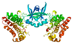
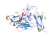
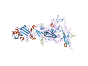
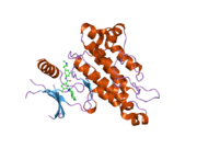
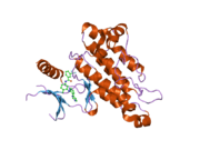
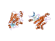
TEK tyrosine kinase
Angiopoietin-1 receptor also known as CD202B (cluster of differentiation 202B) is a protein that in humans is encoded by the TEK gene.[5][6] Also known as TIE2, it is an angiopoietin receptor.
Function
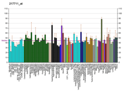
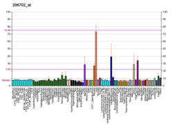
The TEK receptor tyrosine kinase is expressed almost exclusively in endothelial cells in mice, rats, and humans. (TEK is closely related to the TIE receptor tyrosine kinase.)[7]
This receptor possesses a unique extracellular domain containing 2 immunoglobulin-like loops separated by 3 epidermal growth factor-like repeats that are connected to 3 fibronectin type III-like repeats.[8] The ligand for the receptor is angiopoietin-1.[7] TEK has also been suggested as a marker for nucleus pulposus progenitor cells, from the intervertebral disc, which upon activation by Angiopoietin-1 starts to multiply and differentiate.[9][10]
Defects in TEK are associated with inherited venous malformations; the TEK signaling pathway appears to be critical for endothelial cell-smooth muscle cell communication in venous morphogenesis.[7]
Interactions
TEK tyrosine kinase has been shown to interact with:
See also
- Tie-2/Ang-1 signaling
References
- GRCh38: Ensembl release 89: ENSG00000120156 - Ensembl, May 2017
- GRCm38: Ensembl release 89: ENSMUSG00000006386 - Ensembl, May 2017
- "Human PubMed Reference:". National Center for Biotechnology Information, U.S. National Library of Medicine.
- "Mouse PubMed Reference:". National Center for Biotechnology Information, U.S. National Library of Medicine.
- Partanen J, Armstrong E, Mäkelä TP, Korhonen J, Sandberg M, Renkonen R, Knuutila S, Huebner K, Alitalo K (April 1992). "A novel endothelial cell surface receptor tyrosine kinase with extracellular epidermal growth factor homology domains". Molecular and Cellular Biology. 12 (4): 1698–707. doi:10.1128/mcb.12.4.1698. PMC 369613. PMID 1312667.
- Boon LM, Mulliken JB, Vikkula M, Watkins H, Seidman J, Olsen BR, Warman ML (September 1994). "Assignment of a locus for dominantly inherited venous malformations to chromosome 9p". Human Molecular Genetics. 3 (9): 1583–7. doi:10.1093/hmg/3.9.1583. PMID 7833915.
- "Entrez Gene: TEK TEK tyrosine kinase, endothelial (venous malformations, multiple cutaneous and mucosal)".
- Fiedler U, Krissl T, Koidl S, Weiss C, Koblizek T, Deutsch U, Martiny-Baron G, Marmé D, Augustin HG (January 2003). "Angiopoietin-1 and angiopoietin-2 share the same binding domains in the Tie-2 receptor involving the first Ig-like loop and the epidermal growth factor-like repeats". The Journal of Biological Chemistry. 278 (3): 1721–7. doi:10.1074/jbc.M208550200. PMID 12427764.
- Sakai D, Nakamura Y, Nakai T, Mishima T, Kato S, Grad S, Alini M, Risbud MV, Chan D, Cheah KS, Yamamura K, Masuda K, Okano H, Ando K, Mochida J (2012). "Exhaustion of nucleus pulposus progenitor cells with ageing and degeneration of the intervertebral disc". Nature Communications. 3: 1264. Bibcode:2012NatCo...3.1264S. doi:10.1038/ncomms2226. PMC 3535337. PMID 23232394.
- Sakai D, Schol J, Bach FC, Tekari A, Sagawa N, Nakamura Y, Chan SC, Nakai T, Creemers LB (June 2018). "Successful fishing for nucleus pulposus progenitor cells of the intervertebral disc across species". JOR Spine. 1 (2): e1018. doi:10.1002/jsp2.1018. ISSN 2572-1143. PMC 6686801. PMID 31463445.
- Sato A, Iwama A, Takakura N, Nishio H, Yancopoulos GD, Suda T (August 1998). "Characterization of TEK receptor tyrosine kinase and its ligands, Angiopoietins, in human hematopoietic progenitor cells". International Immunology. 10 (8): 1217–27. doi:10.1093/intimm/10.8.1217. PMID 9723709.
- Maisonpierre PC, Suri C, Jones PF, Bartunkova S, Wiegand SJ, Radziejewski C, Compton D, McClain J, Aldrich TH, Papadopoulos N, Daly TJ, Davis S, Sato TN, Yancopoulos GD (July 1997). "Angiopoietin-2, a natural antagonist for Tie2 that disrupts in vivo angiogenesis". Science. 277 (5322): 55–60. doi:10.1126/science.277.5322.55. PMID 9204896.
- Davis S, Aldrich TH, Jones PF, Acheson A, Compton DL, Jain V, Ryan TE, Bruno J, Radziejewski C, Maisonpierre PC, Yancopoulos GD (December 1996). "Isolation of angiopoietin-1, a ligand for the TIE2 receptor, by secretion-trap expression cloning". Cell. 87 (7): 1161–9. doi:10.1016/s0092-8674(00)81812-7. PMID 8980223.
- Jones N, Dumont DJ (September 1998). "The Tek/Tie2 receptor signals through a novel Dok-related docking protein, Dok-R". Oncogene. 17 (9): 1097–108. doi:10.1038/sj.onc.1202115. PMID 9764820.
- Master Z, Jones N, Tran J, Jones J, Kerbel RS, Dumont DJ (November 2001). "Dok-R plays a pivotal role in angiopoietin-1-dependent cell migration through recruitment and activation of Pak". The EMBO Journal. 20 (21): 5919–28. doi:10.1093/emboj/20.21.5919. PMC 125712. PMID 11689432.
Further reading
- Huang L, Turck CW, Rao P, Peters KG (November 1995). "GRB2 and SH-PTP2: potentially important endothelial signaling molecules downstream of the TEK/TIE2 receptor tyrosine kinase". Oncogene. 11 (10): 2097–103. PMID 7478529.
- Deans JP, Kalt L, Ledbetter JA, Schieven GL, Bolen JB, Johnson P (September 1995). "Association of 75/80-kDa phosphoproteins and the tyrosine kinases Lyn, Fyn, and Lck with the B cell molecule CD20. Evidence against involvement of the cytoplasmic regions of CD20". The Journal of Biological Chemistry. 270 (38): 22632–8. doi:10.1074/jbc.270.38.22632. PMID 7545683.
- Gallione CJ, Pasyk KA, Boon LM, Lennon F, Johnson DW, Helmbold EA, Markel DS, Vikkula M, Mulliken JB, Warman ML (March 1995). "A gene for familial venous malformations maps to chromosome 9p in a second large kindred". Journal of Medical Genetics. 32 (3): 197–9. doi:10.1136/jmg.32.3.197. PMC 1050316. PMID 7783168.
- Robertson NG, Khetarpal U, Gutiérrez-Espeleta GA, Bieber FR, Morton CC (September 1994). "Isolation of novel and known genes from a human fetal cochlear cDNA library using subtractive hybridization and differential screening". Genomics. 23 (1): 42–50. doi:10.1006/geno.1994.1457. PMID 7829101.
- Dumont DJ, Anderson L, Breitman ML, Duncan AM (September 1994). "Assignment of the endothelial-specific protein receptor tyrosine kinase gene (TEK) to human chromosome 9p21". Genomics. 23 (2): 512–3. doi:10.1006/geno.1994.1536. PMID 7835909.
- Ziegler SF, Bird TA, Schneringer JA, Schooley KA, Baum PR (March 1993). "Molecular cloning and characterization of a novel receptor protein tyrosine kinase from human placenta". Oncogene. 8 (3): 663–70. PMID 8382358.
- Davis S, Aldrich TH, Jones PF, Acheson A, Compton DL, Jain V, Ryan TE, Bruno J, Radziejewski C, Maisonpierre PC, Yancopoulos GD (December 1996). "Isolation of angiopoietin-1, a ligand for the TIE2 receptor, by secretion-trap expression cloning". Cell. 87 (7): 1161–9. doi:10.1016/S0092-8674(00)81812-7. PMID 8980223.
- Suri C, Jones PF, Patan S, Bartunkova S, Maisonpierre PC, Davis S, Sato TN, Yancopoulos GD (December 1996). "Requisite role of angiopoietin-1, a ligand for the TIE2 receptor, during embryonic angiogenesis". Cell. 87 (7): 1171–80. doi:10.1016/S0092-8674(00)81813-9. PMID 8980224.
- Vikkula M, Boon LM, Carraway KL, Calvert JT, Diamonti AJ, Goumnerov B, Pasyk KA, Marchuk DA, Warman ML, Cantley LC, Mulliken JB, Olsen BR (December 1996). "Vascular dysmorphogenesis caused by an activating mutation in the receptor tyrosine kinase TIE2". Cell. 87 (7): 1181–90. doi:10.1016/S0092-8674(00)81814-0. PMID 8980225.
- Witzenbichler B, Maisonpierre PC, Jones P, Yancopoulos GD, Isner JM (July 1998). "Chemotactic properties of angiopoietin-1 and -2, ligands for the endothelial-specific receptor tyrosine kinase Tie2". The Journal of Biological Chemistry. 273 (29): 18514–21. doi:10.1074/jbc.273.29.18514. PMID 9660821.
- Asahara T, Chen D, Takahashi T, Fujikawa K, Kearney M, Magner M, Yancopoulos GD, Isner JM (August 1998). "Tie2 receptor ligands, angiopoietin-1 and angiopoietin-2, modulate VEGF-induced postnatal neovascularization". Circulation Research. 83 (3): 233–40. doi:10.1161/01.res.83.3.233. PMID 9710115.
- Sato A, Iwama A, Takakura N, Nishio H, Yancopoulos GD, Suda T (August 1998). "Characterization of TEK receptor tyrosine kinase and its ligands, Angiopoietins, in human hematopoietic progenitor cells". International Immunology. 10 (8): 1217–27. doi:10.1093/intimm/10.8.1217. PMID 9723709.
- Jones N, Dumont DJ (September 1998). "The Tek/Tie2 receptor signals through a novel Dok-related docking protein, Dok-R". Oncogene. 17 (9): 1097–108. doi:10.1038/sj.onc.1202115. PMID 9764820.
- De Sepulveda P, Okkenhaug K, Rose JL, Hawley RG, Dubreuil P, Rottapel R (February 1999). "Socs1 binds to multiple signalling proteins and suppresses steel factor-dependent proliferation". The EMBO Journal. 18 (4): 904–15. doi:10.1093/emboj/18.4.904. PMC 1171183. PMID 10022833.
- Valenzuela DM, Griffiths JA, Rojas J, Aldrich TH, Jones PF, Zhou H, McClain J, Copeland NG, Gilbert DJ, Jenkins NA, Huang T, Papadopoulos N, Maisonpierre PC, Davis S, Yancopoulos GD (March 1999). "Angiopoietins 3 and 4: diverging gene counterparts in mice and humans". Proceedings of the National Academy of Sciences of the United States of America. 96 (5): 1904–9. Bibcode:1999PNAS...96.1904V. doi:10.1073/pnas.96.5.1904. PMC 26709. PMID 10051567.
- Calvert JT, Riney TJ, Kontos CD, Cha EH, Prieto VG, Shea CR, Berg JN, Nevin NC, Simpson SA, Pasyk KA, Speer MC, Peters KG, Marchuk DA (July 1999). "Allelic and locus heterogeneity in inherited venous malformations". Human Molecular Genetics. 8 (7): 1279–89. doi:10.1093/hmg/8.7.1279. PMID 10369874.
- Jones N, Master Z, Jones J, Bouchard D, Gunji Y, Sasaki H, Daly R, Alitalo K, Dumont DJ (October 1999). "Identification of Tek/Tie2 binding partners. Binding to a multifunctional docking site mediates cell survival and migration". The Journal of Biological Chemistry. 274 (43): 30896–905. doi:10.1074/jbc.274.43.30896. PMID 10521483.
- Fachinger G, Deutsch U, Risau W (October 1999). "Functional interaction of vascular endothelial-protein-tyrosine phosphatase with the angiopoietin receptor Tie-2". Oncogene. 18 (43): 5948–53. doi:10.1038/sj.onc.1202992. PMID 10557082.
External links
- GeneReviews/NCBI/NIH/UW entry on Multiple Cutaneous and Mucosal Venous Malformations
- Overview of all the structural information available in the PDB for UniProt: Q02763 (Angiopoietin-1 receptor) at the PDBe-KB.
This article incorporates text from the United States National Library of Medicine, which is in the public domain.
