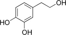
Hydroxytyrosol
Hydroxytyrosol is a phenylethanoid, a type of phenolic phytochemical with antioxidant properties in vitro. In nature, hydroxytyrosol is found in olive leaf and olive oil, in the form of its elenolic acid ester oleuropein and, especially after degradation, in its plain form.[1]
 | |
| Names | |
|---|---|
| IUPAC name
4-(2-Hydroxyethyl)-1,2-benzenediol | |
| Other names
3-Hydroxytyrosol 3,4-dihydroxyphenylethanol (DOPET) Dihydroxyphenylethanol 2-(3,4-Di-hydroxyphenyl)-ethanol (DHPE) 3,4-dihydroxyphenolethanol (3,4-DHPEA)[1] | |
| Identifiers | |
3D model (JSmol) |
|
| ChEBI | |
| ChEMBL | |
| ChemSpider | |
| ECHA InfoCard | 100.114.418 |
PubChem CID |
|
| UNII | |
CompTox Dashboard (EPA) |
|
| |
| |
| Properties | |
| C8H10O3 | |
| Molar mass | 154.165 g·mol−1 |
| Appearance | Clear, faint yellow to yellow liquid |
| Boiling point | 174 °C (345 °F; 447 K) |
| 5 g/100 ml | |
| Hazards | |
| Main hazards | Causes skin irritation.
Causes serious eye irritation. May cause respiratory irritation. |
| Safety data sheet | |
| R-phrases (outdated) | R36/37/38 |
| S-phrases (outdated) | S26, S37/39 |
| Related compounds | |
Related alcohols |
benzyl alcohol, tyrosol |
Except where otherwise noted, data are given for materials in their standard state (at 25 °C [77 °F], 100 kPa). | |
| Infobox references | |
Hydroxytyrosol itself in pure form is a colorless, odorless liquid. The olives, leaves and olive pulp contain large amounts of hydroxytyrosol (compared to olive oil), most of which can be recovered to produce hydroxytyrosol extracts.[1] However, it was found that black olives, such as common canned variety, containing iron(II) gluconate contained little hydroxytyrosol, as iron salts are catalysts for its oxidation.[2]
Hydroxytyrosol is mentioned by the scientific committee of the European Food Safety Authority as one of several olive oil polyphenols under preliminary research for the potential to affect blood lipid levels, although there is no evidence from high-quality clinical research to indicate that this effect exists.[3]
Animal research
As of 2015, the NOAEL for hydroxytyrosol in rats is 250 mg/kg/day, with a LOAEL of 500 mg/kg/day.[4]
Role in neuroprotection and in neurogenesis
In vivo data provide evidence of the neuroprotective effects of oral hydroxytyrosol (HTyr) intake. Both in vivo and in vitro studies have identified mitochondria as one of the targets of the protective effects of hydroxytyrosol in the brain [5][6].
Moreover, a recent study of the effect of hydroxytyrosol treatment in vivo in adult mouse, showed that hydroxytyrosol increases the number of new neurons of the dentate gyrus of the hippocampus (one of the two major neurogenic niches in the brain that continues to produce neurons throughout life) by enhancing their survival without effect on the proliferation of stem and progenitor cells [7]. Notably, however, in aged mice hydroxytyrosol increases not only the survival of new neurons and improves their integration into memory circuits, but also strongly increases the proliferation of stem and progenitor cells and reduces aging markers such as lipofuscin and Iba-1 [7]. Thus, hydroxytyrosol is able to counteract the effect of aging on neurogenesis. Hydroxytyrosol is therefore endowed with pro-neurogenic capability, but, unlike several other neurogenic stimuli (e.g., physical exercise, antidepressants etc., ) has the unique ability to activate stem cells in aging mouse (see for review [8]).
See also
- Echinacoside, a hydroxytyrosol-containing glycoside
- Tyrosol
- Verbascoside, another hydroxytyrosol-containing glycoside
References
- M. Baldioli; M. Servili; G. Perretti; G. F. Montedoro (1996). "Antioxidant activity of tocopherols and phenolic compounds of virgin olive oil". Journal of the American Oil Chemists' Society. 73 (11): 1589–1593. doi:10.1007/BF02523530.
- Vincenzo Marsilio; Cristina Campestre; Barbara Lanza (July 2001). "Phenolic compounds change during California-style ripe olive processing". Food Chemistry. 74 (1): 55–60. doi:10.1016/S0308-8146(00)00338-1.
- Sadler MJ (2014). Foods, Nutrients and Food Ingredients with Authorised EU Health Claims; Volume 1 of Woodhead Publishing Series in Food Science, Technology and Nutrition; Section 10.3: Authorised health claim. Elsevier. pp. 214–5. ISBN 9780857098481.
- Heilman J, Anyangwe N, Tran N, Edwards J, Beilstein P, López J (2015). "Toxicological evaluation of an olive extract, H35: Subchronic toxicity in the rat" (PDF). Food and Chemical Toxicology. 84: 18–28. doi:10.1016/j.fct.2015.07.007. PMID 26184542. Retrieved 2016-05-07.
The lowest observed adverse effect level (LOAEL) was the 500 mg HT/kg bw/day based on statistically significant reductions in body weight gain and decreased body weight in males. The no observed adverse effect level (NOAEL) was 250 mg HT/kg bw/day, equivalent to 691 mg/kg bw/day of H35 extract.
- Schaffer, Sebastian; Podstawa, Maciej; Visioli, Francesco; Bogani, Paola; Müller, Walter E.; Eckert, Gunter P. (2007). "Hydroxytyrosol-Rich Olive Mill Wastewater Extract Protects Brain Cells in Vitro and ex Vivo". Journal of Agricultural and Food Chemistry. 55 (13): 5043–5049. doi:10.1021/jf0703710. ISSN 0021-8561.
- Schaffer, Sebastian; Müller, Walter E.; Eckert, Gunter P. (2010). "Cytoprotective effects of olive mill wastewater extract and its main constituent hydroxytyrosol in PC12 cells". Pharmacological Research. 62 (4): 322–327. doi:10.1016/j.phrs.2010.06.004. ISSN 1043-6618.
- D’Andrea, Giorgio; Ceccarelli, Manuela; Bernini, Roberta; Clemente, Mariangela; Santi, Luca; Caruso, Carla; Micheli, Laura; Tirone, Felice (2020). "Hydroxytyrosol stimulates neurogenesis in aged dentate gyrus by enhancing stem and progenitor cell proliferation and neuron survival". The FASEB Journal. 34 (3): 4512–4526. doi:10.1096/fj.201902643R. ISSN 0892-6638.
- Ceccarelli, Manuela; D’Andrea, Giorgio; Micheli, Laura; Tirone, Felice (2020). "Interaction Between Neurogenic Stimuli and the Gene Network Controlling the Activation of Stem Cells of the Adult Neurogenic Niches, in Physiological and Pathological Conditions". Frontiers in Cell and Developmental Biology. 8. doi:10.3389/fcell.2020.00211. ISSN 2296-634X.