Dieulafoy's lesion
Dieulafoy's lesion is a medical condition characterized by a large tortuous arteriole most commonly in the stomach wall (submucosal) that erodes and bleeds. It can present in any part of the gastrointestinal tract.[1] It can cause gastric hemorrhage[2] but is relatively uncommon. It is thought to cause less than 5% of all gastrointestinal bleeds in adults. It was named after French surgeon Paul Georges Dieulafoy, who described this condition in his paper "Exulceratio simplex: Leçons 1-3" in 1898.[3][4] It is also called "caliber-persistent artery" or "aneurysm" of gastric vessels. However, unlike most other aneurysms, these are thought to be developmental malformations rather than degenerative changes.
| Dieulafoy's lesion | |
|---|---|
| Other names | Exulceratio simplex Dieulafoy |
 | |
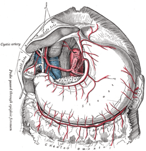
| Blood supply of stomach | |
| Specialty | Gastroenterology |
History
Dieulafoy's lesion was first described in 1884 by M.T. Gallard.[5] The lesion was named after French surgeon Paul Georges Dieulafoy, who described the condition in his paper "Exulceratio simplex: Leçons 1-3" in 1898.[5][3][4] Dieulafoy believed (incorrectly) the bleeding from this lesion was due to erosions of the mucosa in the stomach.[5]
Signs and symptoms
Dieulafoy's lesion often do not cause symptoms (asymptomatic). When present, symptoms usually relate to painless bleeding, with vomiting blood (hematemesis) and/or black stools (melena).[1] Less often, Dieulafoy's lesions may cause rectal bleeding (hematochezia), or rarely, iron deficiency anemia. Usually, there are no gastrointestinal symptoms that precede the bleeding (abdominal pain, nausea, etc.).
| Presenting Symptoms | |
|---|---|
| Recurrent hematemesis with melena | 51% of cases |
| Hematemesis without melena | 28% of cases |
| Melena with no hematemesis | 18% of cases |
Though exceptionally rare, cases of Dieulafoy lesions occurring in the gallbladder can cause upper abdominal pain, which is usually right upper quadrant or upper middle (epigastric).[6] Though gallbladder Dieulafoy lesions usually occur with anemia (83%), they generally do not cause overt bleeding (hematochezia, hematemesis, melena, etc.).[6]
Diagnosis
A Dieulafoy's lesion is difficult to diagnose, because of the intermittent pattern of bleeding. Dieulafoy's lesion are typically diagnose during endoscopic evaluation, usually during upper endoscopy. Lesions affecting the colon or end of the small bowel (terminal ileum) may be diagnosed during colonoscopy. Dieulafoy's lesions are not easily recognized and therefore multiple evaluations with endoscopy may be necessary. Angiography may be helpful with diagnosis, though this only identifies bleeding that actively occurs during the time of that test.
Once identified during endoscopy, the mucosa near a Dieulafoy's lesion may be injected with ink. Tattooing the area can aid in identifying the location of the Dieulafoy's lesion in the event of rebleeding.
Cause
A Dieulafoy's lesion is caused by an abnormally large blood vessel (arteriole) beneath the gastrointestinal mucosa (submucosal) that bleeds, in the absence of any ulcer, erosion, or other abnormality in the mucosa. The size of these blood vessels varies from 1 to 3 mm. It may be that pulsation from the enlarged vessels leads to thinning of the mucosa at that location, leading to exposure of the vessel and subsequent hemorrhage.
In contrast to peptic ulcer disease, a history of alcohol abuse or NSAID use is usually absent in Dieulafoy's lesion. It can present in any part of the gastrointestinal tract.[1] Dieulafoy's lesions have been reported in the gallbladder.
Pathophysiology
Dieulafoy's lesions are characterized by a single large tortuous small artery[7] in the submucosa which does not undergo normal branching or a branch with caliber of 1–5 mm (more than 10 times the normal diameter of mucosal capillaries). The lesion bleeds into the gastrointestinal tract through a minute defect in the mucosa which is not a primary ulcer of the mucosa but an erosion likely caused in the submucosal surface by protrusion of the pulsatile arteriole.
Approximately 75% of Dieulafoy's lesions occur in the upper part of the stomach within 6 cm of the gastroesophageal junction, most commonly in the lesser curvature. Extragastric lesions have historically been thought to be uncommon but have been identified more frequently in recent years, likely due to increased awareness of the condition. The duodenum is the most common location (14%) followed by the colon (5%), surgical anastamoses (5%), the jejunum (1%) and the esophagus (1%).[8] The pathology in these extragastric locations is essentially the same as that of the more common gastric lesion.
Treatment
It is diagnosed and treated endoscopically; however, endoscopic ultrasound or angiography can be of benefit.
Endoscopic techniques used in the treatment include epinephrine injection followed by bipolar or monopolar electrocoagulation, injection sclerotherapy, heater probe, laser photocoagulation, hemoclipping or banding. In cases of refractory bleeding, interventional radiology may be consulted for an angiogram with subselective embolization.[9]
Prognosis
The mortality rate for Dieulafoy's was much higher before the era of endoscopy, where open surgery was the only treatment option. Long term control of bleeding (hemostasis) is achieved in 85 - 90 percent of cases.
Epidemiology
Dieulafoy's lesions account for roughly 1.5 percent of gastrointestinal hemorrhage.[5] These lesions are twice as common in men, and often occur in older individuals (over 50 years of age) with multiple comorbidities, including hypertension, cardiovascular disease, chronic kidney disease, and diabetes.
References
- al-Mishlab T, Amin AM, Ellul JP (August 1999). "Dieulafoy's lesion: an obscure cause of GI bleeding". Journal of the Royal College of Surgeons of Edinburgh. 44 (3): 222–5. PMID 10453143.
- Akhras J, Patel P, Tobi M (March 2007). "Dieulafoy's lesion-like bleeding: an underrecognized cause of upper gastrointestinal hemorrhage in patients with advanced liver disease". Dig. Dis. Sci. 52 (3): 722–6. doi:10.1007/s10620-006-9468-7. PMID 17237996.
- synd/3117 at Who Named It?
- G. Dieulafoy. Exulceratio simplex: Leçons 1-3. In: G. Dieulafoy, editor: Clinique medicale de l'Hotel Dieu de Paris. Paris, Masson et Cie: 1898:1-38.
- Inayat, F; Ullah, W; Hussain, Q; Hurairah, A (6 January 2017). "Dieulafoy's lesion of the oesophagus: a case series and literature review". BMJ Case Reports. 2017: bcr2016218100. doi:10.1136/bcr-2016-218100. PMC 5256583. PMID 28062437.
- Wu, JM; Zaitoun, AM (2018). "A galling disease? Dieulafoy's lesion of the gallbladder". International Journal of Surgery Case Reports. 44: 62–65. doi:10.1016/j.ijscr.2018.01.027. PMC 5928034. PMID 29477106.
- Eidus, LB.; Rasuli, P.; Manion, D.; Heringer, R. (Nov 1990). "Caliber-persistent artery of the stomach (Dieulafoy's vascular malformation)". Gastroenterology. 99 (5): 1507–10. doi:10.1016/0016-5085(90)91183-7. PMID 2210260.
- Lee Y, Walmsley R, Leong R, Sung J (2003). "Dieulafoy's Lesion". Gastrointestinal Endoscopy. 58 (2): 236–243. doi:10.1067/mge.2003.328. PMID 12872092.
- Navuluri, Rakesh; Kang, Lisa; Patel, Jay; Van Ha, Thuong (2012-09-01). "Acute Lower Gastrointestinal Bleeding". Seminars in Interventional Radiology. 29 (3): 178–186. doi:10.1055/s-0032-1326926. ISSN 0739-9529. PMC 3577586. PMID 23997409.