Otocephaly
Otocephaly, also known as agnathia–otocephaly complex, is a very rare and lethal cephalic disorder characterized by the absence of the mandible (agnathia), with the ears fused together just below the chin (synotia). It is caused by a disruption to the development of the first branchial arch. It occurs in every 1 in 70,000 embryos.
| Otocephaly | |
|---|---|
| Other names | Agnathia-otocephaly complex,[1] dysgnathia complex,[1] holoprosencephaly–agnathia,[1] Kanwar syndrome[2] |
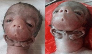 | |
| Female infant with otocephaly | |
| Specialty | Medical genetics |
| Symptoms | Absence of mandible (agnathia), small or absent mouth (microstomia), fused ears below chin (synotia), holoprosencephaly |
| Usual onset | 23rd–26th day of gestation (Carnegie stage 10) |
| Causes | Genetic |
| Diagnostic method | Prenatal ultrasound |
| Differential diagnosis | Treacher Collins syndrome, Goldenhar syndrome, Möbius syndrome |
| Prognosis | Stillbirth or miscarriage |
| Frequency | 1:70,000 |
Signs and symptoms
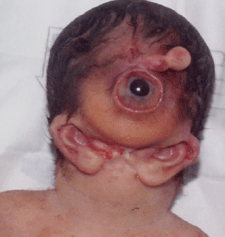
The disorder is characterised by the absence of the mandible (agnathia), with the ears fused together just below the chin (synotia). In addition to agnathia and synotia, other symptoms that may manifest in otocephaly include:[2]
- Facial/musculoskeletal
- Small (microglossia) or absent (aglossia) tongue
- Small (microstomia) or absent (astomia) mouth
- Cyclopia with proboscis
- Median cleft lip and cleft palate
- Neurological
- Organ systems
- Situs inversus (full rotation of the internal organs)
- Cardiac anomalies
- Ambiguous genitalia
- Absence of glands
Grades
Sewall Wright described twelve grades of otocephaly.[3] Grades 1 to 5 were isolated agnathia with no neurological defects. Grades 6 to 9 were cyclopia with holoprosencephaly. Grades 10 to 12 were aprosopus with absence of the prosencephalon and mesencephalon. In aprosopus, the face is entirely absent from the cranium, and one human case of aprosopus has been reported in modern history.[4]
Cause
Otocephaly is generally a result of a de novo mutation. Mutations in the gene PRRX1 on the long arm of chromosome 1 have been in some medical cases.[1] Autosomal trisomies, while prevalent in similar conditions like cyclopia, are uncommon in otocephaly.
Development
During early embryogenesis, many different organ systems begin development. Any disruption in these processes results in complex malformation that usually results in death. The first branchial arch will normally develop around the 23rd to 26th day of gestation, also known as Carnegie stage 10. Usually, failure of this will result in isolated agnathia, but otocephaly may occur in exceptional circumstances. After agenesis of the first branchial arch, no cure is possible.
Prognosis
In almost all cases, fetuses with otocephaly are naturally or electively aborted before birth. If the infant is carried to term, death occurs within minutes due to airway obstruction.
History
Otocephaly was first described in 1717 by Dutch scientist Theodor Kerckring. In 1933, evolutionary biologist Sewall Wright performed a study on otocephaly on guinea pigs and gave otocephaly its name.[3] The name comes from the Greek-derived New Latin prefix oto- ("ear") and the suffix -cephaly ("head").
In 2018, Indian neonatologist Kanwar Singh and his associates described a severe case of otocephaly with cyclopia, agnathia, complete astomia and synotia. They termed this severe presentation Kanwar syndrome.[2]
References
- "OMIM Entry - # 202650 - AGNATHIA-OTOCEPHALY COMPLEX; AGOTC". Online Mendelian Inheritance in Man. Retrieved 2019-06-05.
- Singh, K; Sharma, S; Agarwal, K; Kalra, A (2018). "Cyclopia-otocephaly-agnathia-synotia-astomia complex: A case report". Journal of Clinical Neonatology. 7 (3): 177. doi:10.4103/jcn.jcn_23_18. ISSN 2249-4847.
- Wright, S (Nov 1934). "On the Genetics of Subnormal Development of the Head (Otocephaly) in the Guinea Pig". Genetics. 19 (6): 471–505. PMC 1208510. PMID 17246734.
- Utkus, A; Kazakevicius, R; Ptasekas, R; Kucinskas, V; Beckwith, JB; Opitz, JM (2001). "Human anotocephaly (aprosopus, acrania-synotia) in the Vilnius anatomical collection". American Journal of Medical Genetics. 101 (2): 163–171. doi:10.1002/ajmg.1320. PMID 11391661.