NUMB (gene)
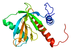
Protein numb homolog is a protein that in humans is encoded by the NUMB gene. The protein encoded by this gene plays a role in the determination of cell fates during development. The encoded protein, whose degradation is induced in a proteasome-dependent manner by MDM2, is a membrane-bound protein that has been shown to associate with EPS15, LNX1, and NOTCH1. Four transcript variants encoding different isoforms have been found for this gene.[5]
The protein Numb is coded for by the gene, NUMB, whose mechanism appears to be evolutionarily conserved.[6] Numb has been extensively studied in both invertebrates and mammals, though its function is best understood in Drosophila. Numb plays a crucial role in asymmetrical cell division during development, allowing for differential cell fate specification in the central and peripheral nervous systems. During neurogenesis, Numb localizes to one side of the mother cell such that it is distributed selectively to one daughter cell. This asymmetric division allows a daughter cell containing Numb to acquire a different fate than the other daughter cell.
Gene
The numb gene protein product controls binary cell fate decisions in the peripheral and central nervous systems of both invertebrates and mammals during neurogenesis.[7] During cell division, Numb is asymmetrically localized to one end of the progenitor cell and subsequently segregates to only one daughter cell where it intrinsically determines cell fate.[7] Numb protein signaling plays a key role in binary cell fate decisions following asymmetric cell divisions. One daughter cell, generally that receiving the Numb, is able to adopt a neuronal fate and innervate the developing nervous system. The other daughter cell becomes a progenitor cell to fill the lost role of the parent cell and maintain proliferation. In addition to its role in proliferation and differentiation, Numb has also been shown to play a role in tumorigenesis and the response of neural progenitors to chemotactic cues during migration.
In mammals, there are four alternatively spliced forms of the Numb protein. In addition, there is a Numb homolog called “Numb-like,” or NUMBL. Numb proteins in mammals are not as well understood as their fly counterparts. The various forms of Numb have differential progenitor-promoting and differentiation-promoting functions.[8] More research is necessary to understand the complex relationships between these forms of Numb and their functions.
Asymmetic localization
In both invertebrates and mammals, Numb is localized using the Pins/GαI complex and the PAR complex of Bazooka (Par3 in mammals), Par6, and aPKC (atypical protein kinase C). In the sensory organ precursor (SOP) cell, the PAR proteins localize to the posterior pole of the cell, and the Pins/GαI complex is localized to the anterior pole of the cell. This results in an anterior/posterior cell division with daughter cells of similar size. In neuroblasts, both complexes are localized to the apical cortex, causing apical/basal cell division and daughter cells exhibiting strong size asymmetry.[9] In the SOP, one mechanism for Numb localization has been proposed based on the PAR complex. It states that a complex phosphorylation cascade enables aPKC to phosphorylate Numb in the pre-mitotic cell, decreasing its affinity for the plasma membrane. This releases Numb from the aPKC pole, increasing its presence in the non-aPKC pole.[10] This establishes the asymmetric distribution of Numb, with the Numb/Pon crescent on one side of the mother cell.
Another proposed component of the localization complex is Partner of Numb (PON), which is asymmetrically localized during mitosis and acts as an adaptor protein by binding and mediating the anchoring of Numb. The localization of PON is controlled by either Insc or the Frizzled-Wnt signaling pathway.[11]
Role in cell proliferation and differentiation
Differentiation through inhibition of notch signaling
Numb’s primary function in cell differentiation is as an inhibitor of Notch signaling which is essential for maintaining self-renewal potential in stem and progenitor cells. Notch is a transmembrane signaling receptor that is activated by DSL family ligands. Notch binds the ligands Delta and Serrate in Drosophila. The human ligands are Delta-like and Jagged, respectively. These ligands are themselves integral membrane proteins. Following ligand binding of the Notch receptor, the intracellular fragment of Notch (NICD, or notch intracellular domain) is released into the cytoplasm and transported to the nucleus, where it can form a complex with binding partners such as EP300 and histone acetyltransferase and act as a transcription factor for Notch target genes.[12] Among the Notch target genes are members of the HES and HEY gene families whose protein products can act as transcriptional repressors for tissue-specific transcription factors, thus maintaining the cell’s potential for self-renewal.
Inhibition of notch signaling through the ubiquitination pathway
Numb exerts its functional role on cell fate decisions by antagonizing Notch signaling activities. The molecular mechanisms underlying this relationship appear to rely on the ubiquitination of the membrane bound Notch1 receptor and the subsequent degradation of its NICD following receptor activation.[13] In support of this, Numb’s ability to ubiquinate Notch1 directly correlated with its functional inhibition of Notch1 signaling activities. The ubiquitination pathway directs protein recycling by directly tagging specific proteins for proteasome degradation. Through a multi-step process, free ubiquitin is first attached to an activating enzyme (E1) and then transferred to a conjugating enzyme (E2) which partners with a ligase (E3) which functions as an adaptor to selectively transfer the ubiquitin to specific protein substrates. Numb expression was found to selectively tag the membrane Notch1 receptor for ubiquitination through the interaction of its Phosphotyrosine-binding domain with the E3 ubiquitin ligase Itch. Numb and Itch work in concert to promote the ubiquitination of the full-length membrane-tethered Notch receptor prior to activation. However, Numb only appears to promote the degradation of the NICD cleavage product following receptor activation, targeting it for proteasome degradation and preventing its translocation to the nucleus.
Inhibition of notch signaling through sanpodo
Numb acts as an antagonist for Notch by causing its selective endocytosis and degradation.[14] Another mechanism proposed for how this is accomplished in Drosophila involves a protein called Sanpodo. Sanpodo is a protein that associates with both Notch and Numb. It is located at the plasma membrane and is necessary for Notch activation, promoting Notch cleavage and NICD signaling in the nucleus.[9] Numb converts Sanpodo from an activator to an inhibitor of Notch signaling, magnifying the differences in Notch signaling between different daughter cells. In daughter cells containing Numb, Sanpodo allows Numb to inhibit Notch. In daughter cells with no Numb, Sanpodo potentiates Notch signaling. Sanpodo therefore allows cells to maintain Notch signaling at a below- or above-threshold level.[6]
Numb in Drosophila
Numb has been studied most extensively in Drosophila, in particular in the context of their sensory organ precursors and ganglion mother cells.
External sensory organ development
The Drosophila external sensory organ is a sensory structure in the peripheral nervous system that consists of four cells; a neuron, a sheath cell that surrounds the dendrite, and hair and socket cells, which are considered the “outer” support cells. All four cell fates are descendants of the sensory organ precursor (SOP) cell. In response to the proper cues, SOPs first divide into two secondary precursor cells. The posterior daughter cell is called the pIIa cell and the anterior daughter cell is called the pIIb. The pIIa cell divides to produce a bristle cell and a socket cell, while the pIIb cell divides to produce a neuron and a glial cell. The asymmetric division of the SOP into daughter cells with distinct fates is dependent upon the distribution of Numb. Numb is distributed uniformly in the cytoplasm until mitotic division, when it is selectively localized to the anterior pole of the cell. Thus, Numb is selectively segregated into the pIIb daughter cell upon division of the SOP.[15]
Loss of Numb function causes inappropriate differentiation of SOP cells into all pIIa cells, producing four outer support cells and no neurons or glia.[16] In SOP loss of function Numb mutants, flies have a significant decrease in sensory neurons, leaving them “numb.” Gain of function Notch mutants express a similar phenotype.[17] Ectopic expression of Numb during SOP division has the opposite effect, producing all pIIb cells and no outer support cells. In support of previous experiments demonstrating the role of Numb in Notch signaling inhibition, functional loss of Notch signaling components result in SOP division into two pIIb cells, suggesting Numb promotes acquisition of the pIIb cell fate through inhibition of Notch signaling.[16] Thus, the asymmetric distribution of Numb into IIb secondary precursors during SOP division is necessary for daughter cells to acquire distinct cell fates.[15]
Ganglion mother cell
A ganglion mother cell (GMC) is the cell derived from the division of a neuroblast in the Drosophila central nervous system. The neuroblast divides to produce two cells, a progenitor cell like the mother neuroblast and a GMC that will divide to produce neurons. The mother neuroblast divides along the apical-basal axis, with Numb localizing basally and ending up in the GMC.[18]
Numb in mammals
Alternative splicing to support proliferation and differentiation
In mice embryos mutant for Numb, early neurons emerge in the expected spatial and temporal pattern but fail to maintain a sufficient pool of proliferating progenitors and nearly deplete the population of dividing cells shortly after the onset of neurogenesis.[19] These embryos display precocious neuron production in the forebrain and defects in neural tube closure, dying around embryonic day 11.5.[20] These studies suggest a functional role of mammalian Numb in promoting progenitor cell fate during neurogenesis, which directly opposes the proposed role of Numb in invertebrates. However, other studies have shown overexpression of Numb in the mammalian neural crest MONC-1 stem cell line biases neuronal differentiation, consistent with what is observed in drosophila.[21]
Unlike the invertebrate Numb gene, the mammalian Numb gene undergoes alternative splicing to produce at least four functionally different Numb isoforms. While asymmetric divisions alone can produce sufficient neuron populations in Drosophilia, mammalian brains are much more advanced and require larger populations of neurons that can’t be established to asymmetric divisions alone.[22] Thus, mammalian cortical progenitors must first need to undergo symmetric divisions to expand the progenitor pool before they can undergo later asymmetric divisions for neuronal generations. The mammalian brain has accounted for this by producing isoforms of Numb that maintain progenitor populations in addition to those which support neuronal differentiation.
Studies using the mouse embryonic P19 cell line have shown isoforms with the short proline rich region (PRR) domain promote neuronal differentiation, while those with the long PRR domain promote cell proliferation and prevent differentiation.[23] The p71 and p72 isoforms, which contain the PRR insert, are primarily expressed in actively dividing tissues and are down regulated during differentiation suggesting these isoforms promote cell proliferation (Dho et al., 1999). In contrast, the Drosophilia Numb gene encodes a 66 kDa protein.[21] Consistent with the finding that Numb only supports differentiation and not proliferation in asymmetric division, the 66 kDa Drosophilia protein is analogous to a shorter mammalian isoform lacking the PRR insert and thus promoting cell differentiation.[21]
Role in cancer and tumorigenesis
In several types of cancer, a loss of Numb expression has been demonstrated. This is well-established in breast cancer, where a loss of Numb correlates with a worse prognosis.[24] Numb loss has also been demonstrated in Non-small-cell lung carcinoma, salivary gland carcinoma, and chronic myelogenous leukemia. Restoration of Numb function, or manipulation of enzymes in the ubiquitin mechanism, are some possible research directions for the treatment of certain cancer types.[6]
Role in mammary carcinomas
In approximately half of all human mammary carcinomas, Numb-mediated suppression of Notch signaling is lost due to Numb ubiquitination, tagging it for proteasomal degradation.[24] Numb acts as an oncogene suppressor, inhibiting tumor cell proliferation through suppression of Notch signaling. Increased Notch signaling is observed in tumors where Numb activity has been lost and retrovirally mediated transient overexpression of Numb protein in these tumors restored basal levels of Notch signaling and significantly reduced their colony-forming abilities. Thus, the biological antagonism between Notch and Numb signaling that controls the proliferative/differentiative balance of many cell lineages appears to play a role in human breast carcinogenesis and perhaps other types of tumorigenesis. Pharmalogical inhibition of Notch signaling or enhancement of Numb signaling could be a source of treatment for cancer patients in the future.
Numb has been posited to have a role in tumor-suppression, through its ability to regulate Notch and TP53. Numb binds and inhibits the E3-ligase Mdm2 that is responsible for TP53 ubiquitination and degradation. The ablation of Numb in a cell leads to a decrease in TP53, causing impaired apoptosis and cell cycle checkpoint response. Restoring Numb levels also restores TP53 expression and tumor suppressant abilities.[6]
Role in cell migration
Neural precursors are generated in proliferative zones, before migrating to directed locations where they undergo maturation and become functional neurons. Studies in Drosophilia first suggested Numb played a role in cell migration when mutants displayed defective glial migration along axonal tracts. Since then, a mechanism has been discovered through which Numb binds chemotactic signaling receptors, forming a scaffold for atypical PKC (aPKC) recruitment to the receptor complex.[25] Once activated, aPKC phosphorylates Numb, thus promoting a positive feed-forward response that potentiates Numb-chemotactic receptor binding and subsequent endosomal complex formation. Endocytosis supports the relocalization of the chemotactic receptor to the front of the cell to promote receptor-mediated directional migration in response to receptor activation.
Brain-derived neurotrophic factor is among the chemotactic factors that stimulate Numb-mediated chemotaxis during cell migration.[25] BDNF can function as a chemotactic factor for neural precursors during migration by activating TrkB receptors. Numb binds to TrkB receptors to act as an endocytic regulator of TrkB and promote aPKC activation by acting as a scaffolding protein. Once phosphorylated, aPKC can also phosphorylate Numb to increase its efficacy for binding TrkB, thus promoting the precursor’s chemotactic sensitivity to BDNF.
Interactions
Numb has demonstrated protein-protein interactions with adaptor-related protein complex 2, alpha 1,[26] Mdm2,[27][28] L1,[26] DPYSL2,[26] SIAH1,[29] P53[28] and LNX1.[30]
References
- GRCh38: Ensembl release 89: ENSG00000133961 - Ensembl, May 2017
- GRCm38: Ensembl release 89: ENSMUSG00000021224 - Ensembl, May 2017
- "Human PubMed Reference:". National Center for Biotechnology Information, U.S. National Library of Medicine.
- "Mouse PubMed Reference:". National Center for Biotechnology Information, U.S. National Library of Medicine.
- "Entrez Gene: NUMB numb homolog (Drosophila)".
- Pece S, Confalonieri S, R Romano P, Di Fiore PP (January 2011). "NUMB-ing down cancer by more than just a NOTCH". Biochim. Biophys. Acta. 1815 (1): 26–43. doi:10.1016/j.bbcan.2010.10.001. PMID 20940030.
- Dho SE, French MB, Woods SA, McGlade CJ (November 1999). "Characterization of four mammalian numb protein isoforms. Identification of cytoplasmic and membrane-associated variants of the phosphotyrosine binding domain". J. Biol. Chem. 274 (46): 33097–104. doi:10.1074/jbc.274.46.33097. PMID 10551880.
- Gulino A, Di Marcotullio L, Screpanti I (April 2010). "The multiple functions of Numb". Exp. Cell Res. 316 (6): 900–6. doi:10.1016/j.yexcr.2009.11.017. PMID 19944684.
- Roegiers F, Jan YN (April 2004). "Asymmetric cell division". Curr. Opin. Cell Biol. 16 (2): 195–205. doi:10.1016/j.ceb.2004.02.010. PMID 15196564.
- Wirtz-Peitz F, Nishimura T, Knoblich JA (October 2008). "Linking cell cycle to asymmetric division: Aurora-A phosphorylates the Par complex to regulate Numb localization". Cell. 135 (1): 161–73. doi:10.1016/j.cell.2008.07.049. PMC 2989779. PMID 18854163.
- Lu B, Rothenberg M, Jan LY, Jan YN (October 1998). "Partner of Numb colocalizes with Numb during mitosis and directs Numb asymmetric localization in Drosophila neural and muscle progenitors". Cell. 95 (2): 225–35. doi:10.1016/S0092-8674(00)81753-5. PMID 9790529.
- Katoh M, Katoh M (September 2006). "NUMB is a break of WNT-Notch signaling cycle". Int. J. Mol. Med. 18 (3): 517–21. doi:10.3892/ijmm.18.3.517. PMID 16865239.
- McGill MA, McGlade CJ (June 2003). "Mammalian numb proteins promote Notch1 receptor ubiquitination and degradation of the Notch1 intracellular domain". J. Biol. Chem. 278 (25): 23196–203. doi:10.1074/jbc.M302827200. PMID 12682059.
- Berdnik D, Török T, González-Gaitán M, Knoblich JA (August 2002). "The endocytic protein alpha-Adaptin is required for numb-mediated asymmetric cell division in Drosophila". Dev. Cell. 3 (2): 221–31. doi:10.1016/S1534-5807(02)00215-0. PMID 12194853.
- Rhyu MS, Jan LY, Jan YN (February 1994). "Asymmetric distribution of numb protein during division of the sensory organ precursor cell confers distinct fates to daughter cells". Cell. 76 (3): 477–91. doi:10.1016/0092-8674(94)90112-0. PMID 8313469.
- Spana EP, Doe CQ (July 1996). "Numb antagonizes Notch signaling to specify sibling neuron cell fates". Neuron. 17 (1): 21–6. doi:10.1016/S0896-6273(00)80277-9. PMID 8755475.
- Guo M, Jan LY, Jan YN (July 1996). "Control of daughter cell fates during asymmetric division: interaction of Numb and Notch". Neuron. 17 (1): 27–41. doi:10.1016/S0896-6273(00)80278-0. PMID 8755476.
- Karcavich RE (March 2005). "Generating neuronal diversity in the Drosophila central nervous system: a view from the ganglion mother cells". Dev. Dyn. 232 (3): 609–16. doi:10.1002/dvdy.20273. PMID 15704126.
- Petersen PH, Zou K, Hwang JK, Jan YN, Zhong W (October 2002). "Progenitor cell maintenance requires numb and numblike during mouse neurogenesis". Nature. 419 (6910): 929–34. doi:10.1038/nature01124. PMID 12410312.
- Zhong W, Jiang MM, Schonemann MD, Meneses JJ, Pedersen RA, Jan LY, Jan YN (June 2000). "Mouse numb is an essential gene involved in cortical neurogenesis". Proc. Natl. Acad. Sci. U.S.A. 97 (12): 6844–9. doi:10.1073/pnas.97.12.6844. PMC 18761. PMID 10841580.
- Verdi JM, Bashirullah A, Goldhawk DE, Kubu CJ, Jamali M, Meakin SO, Lipshitz HD (August 1999). "Distinct human NUMB isoforms regulate differentiation vs. proliferation in the neuronal lineage". Proc. Natl. Acad. Sci. U.S.A. 96 (18): 10472–6. doi:10.1073/pnas.96.18.10472. PMC 17913. PMID 10468633.
- Zhong W, Feder JN, Jiang MM, Jan LY, Jan YN (July 1996). "Asymmetric localization of a mammalian numb homolog during mouse cortical neurogenesis". Neuron. 17 (1): 43–53. doi:10.1016/S0896-6273(00)80279-2. PMID 8755477.
- Verdi JM, Schmandt R, Bashirullah A, Jacob S, Salvino R, Craig CG, Program AE, Lipshitz HD, McGlade CJ (September 1996). "Mammalian NUMB is an evolutionarily conserved signaling adapter protein that specifies cell fate". Curr. Biol. 6 (9): 1134–45. doi:10.1016/S0960-9822(02)70680-5. PMID 8805372.
- Pece S, Serresi M, Santolini E, Capra M, Hulleman E, Galimberti V, Zurrida S, Maisonneuve P, Viale G, Di Fiore PP (October 2004). "Loss of negative regulation by Numb over Notch is relevant to human breast carcinogenesis". J. Cell Biol. 167 (2): 215–21. doi:10.1083/jcb.200406140. PMC 2172557. PMID 15492044.
- Zhou P, Alfaro J, Chang EH, Zhao X, Porcionatto M, Segal RA (May 2011). "Numb links extracellular cues to intracellular polarity machinery to promote chemotaxis". Dev. Cell. 20 (5): 610–22. doi:10.1016/j.devcel.2011.04.006. PMC 3103748. PMID 21571219.
- Nishimura T, Fukata Y, Kato K, Yamaguchi T, Matsuura Y, Kamiguchi H, Kaibuchi K (September 2003). "CRMP-2 regulates polarized Numb-mediated endocytosis for axon growth". Nat. Cell Biol. 5 (9): 819–26. doi:10.1038/ncb1039. PMID 12942088.
- Yogosawa S, Miyauchi Y, Honda R, Tanaka H, Yasuda H (March 2003). "Mammalian Numb is a target protein of Mdm2, ubiquitin ligase". Biochem. Biophys. Res. Commun. 302 (4): 869–72. doi:10.1016/S0006-291X(03)00282-1. PMID 12646252.
- Colaluca IN, Tosoni D, Nuciforo P, Senic-Matuglia F, Galimberti V, Viale G, Pece S, Di Fiore PP (January 2008). "NUMB controls p53 tumour suppressor activity". Nature. 451 (7174): 76–80. doi:10.1038/nature06412. PMID 18172499.
- Susini L, Passer BJ, Amzallag-Elbaz N, Juven-Gershon T, Prieur S, Privat N, Tuynder M, Gendron MC, Israël A, Amson R, Oren M, Telerman A (December 2001). "Siah-1 binds and regulates the function of Numb". Proc. Natl. Acad. Sci. U.S.A. 98 (26): 15067–72. doi:10.1073/pnas.261571998. PMC 64984. PMID 11752454.
- Nie J, McGill MA, Dermer M, Dho SE, Wolting CD, McGlade CJ (January 2002). "LNX functions as a RING type E3 ubiquitin ligase that targets the cell fate determinant Numb for ubiquitin-dependent degradation". EMBO J. 21 (1–2): 93–102. doi:10.1093/emboj/21.1.93. PMC 125803. PMID 11782429.
Further reading
- Wong WT, Schumacher C, Salcini AE, et al. (1995). "A protein-binding domain, EH, identified in the receptor tyrosine kinase substrate Eps15 and conserved in evolution". Proc. Natl. Acad. Sci. U.S.A. 92 (21): 9530–4. doi:10.1073/pnas.92.21.9530. PMC 40835. PMID 7568168.
- Sherrington R, Rogaev EI, Liang Y, et al. (1995). "Cloning of a gene bearing missense mutations in early-onset familial Alzheimer's disease". Nature. 375 (6534): 754–60. doi:10.1038/375754a0. PMID 7596406.
- Zhong W, Feder JN, Jiang MM, et al. (1996). "Asymmetric localization of a mammalian numb homolog during mouse cortical neurogenesis". Neuron. 17 (1): 43–53. doi:10.1016/S0896-6273(00)80279-2. PMID 8755477.
- Salcini AE, Confalonieri S, Doria M, et al. (1997). "Binding specificity and in vivo targets of the EH domain, a novel protein–protein interaction module". Genes Dev. 11 (17): 2239–49. doi:10.1101/gad.11.17.2239. PMC 275390. PMID 9303539.
- Dho SE, Jacob S, Wolting CD, et al. (1998). "The mammalian numb phosphotyrosine-binding domain. Characterization of binding specificity and identification of a novel PDZ domain-containing numb binding protein, LNX". J. Biol. Chem. 273 (15): 9179–87. doi:10.1074/jbc.273.15.9179. PMID 9535908.
- Juven-Gershon T, Shifman O, Unger T, et al. (1998). "The Mdm2 Oncoprotein Interacts with the Cell Fate Regulator Numb". Mol. Cell. Biol. 18 (7): 3974–82. PMC 108982. PMID 9632782.
- Santolini E, Puri C, Salcini AE, et al. (2001). "Numb Is an Endocytic Protein". J. Cell Biol. 151 (6): 1345–52. doi:10.1083/jcb.151.6.1345. PMC 2190585. PMID 11121447.
- Susini L; Passer BJ; Amzallag-Elbaz N; et al. (2002). "Siah-1 binds and regulates the function of Numb". Proc. Natl. Acad. Sci. U.S.A. 98 (26): 15067–72. doi:10.1073/pnas.261571998. PMC 64984. PMID 11752454.
- Nie J, McGill MA, Dermer M, et al. (2002). "LNX functions as a RING type E3 ubiquitin ligase that targets the cell fate determinant Numb for ubiquitin-dependent degradation". EMBO J. 21 (1–2): 93–102. doi:10.1093/emboj/21.1.93. PMC 125803. PMID 11782429.
- Rice DS, Northcutt GM, Kurschner C (2002). "The Lnx family proteins function as molecular scaffolds for Numb family proteins". Mol. Cell. Neurosci. 18 (5): 525–40. doi:10.1006/mcne.2001.1024. PMID 11922143.
- Roncarati R, Sestan N, Scheinfeld MH, et al. (2002). "The γ-secretase-generated intracellular domain of β-amyloid precursor protein binds Numb and inhibits Notch signaling". Proc. Natl. Acad. Sci. U.S.A. 99 (10): 7102–7. doi:10.1073/pnas.102192599. PMC 124535. PMID 12011466.
- Strausberg RL, Feingold EA, Grouse LH, et al. (2003). "Generation and initial analysis of more than 15,000 full-length human and mouse cDNA sequences". Proc. Natl. Acad. Sci. U.S.A. 99 (26): 16899–903. doi:10.1073/pnas.242603899. PMC 139241. PMID 12477932.
- Calderwood DA, Fujioka Y, de Pereda JM, et al. (2003). "Integrin β cytoplasmic domain interactions with phosphotyrosine-binding domains: A structural prototype for diversity in integrin signaling". Proc. Natl. Acad. Sci. U.S.A. 100 (5): 2272–7. doi:10.1073/pnas.262791999. PMC 151330. PMID 12606711.
- Yogosawa S, Miyauchi Y, Honda R, et al. (2003). "Mammalian Numb is a target protein of Mdm2, ubiquitin ligase". Biochem. Biophys. Res. Commun. 302 (4): 869–72. doi:10.1016/S0006-291X(03)00282-1. PMID 12646252.
- McGill MA, McGlade CJ (2003). "Mammalian numb proteins promote Notch1 receptor ubiquitination and degradation of the Notch1 intracellular domain". J. Biol. Chem. 278 (25): 23196–203. doi:10.1074/jbc.M302827200. PMID 12682059.
- Rossé C, L'Hoste S, Offner N, et al. (2003). "RLIP, an effector of the Ral GTPases, is a platform for Cdk1 to phosphorylate epsin during the switch off of endocytosis in mitosis". J. Biol. Chem. 278 (33): 30597–604. doi:10.1074/jbc.M302191200. PMID 12775724.
- Nishimura T, Fukata Y, Kato K, et al. (2003). "CRMP-2 regulates polarized Numb-mediated endocytosis for axon growth". Nat. Cell Biol. 5 (9): 819–26. doi:10.1038/ncb1039. PMID 12942088.
- Qin H, Percival-Smith A, Li C, et al. (2004). "A novel transmembrane protein recruits numb to the plasma membrane during asymmetric cell division". J. Biol. Chem. 279 (12): 11304–12. doi:10.1074/jbc.M311733200. PMID 14670962.
- Ota T, Suzuki Y, Nishikawa T, et al. (2004). "Complete sequencing and characterization of 21,243 full-length human cDNAs". Nat. Genet. 36 (1): 40–5. doi:10.1038/ng1285. PMID 14702039.


