Dermatitis
Dermatitis, also known as eczema, is a group of diseases that result in inflammation of the skin.[1] These diseases are characterized by itchiness, red skin and a rash.[1] In cases of short duration, there may be small blisters, while in long-term cases the skin may become thickened.[1] The area of skin involved can vary from small to covering the entire body.[1][2]
| Dermatitis | |
|---|---|
| Other names | Eczema |
 | |
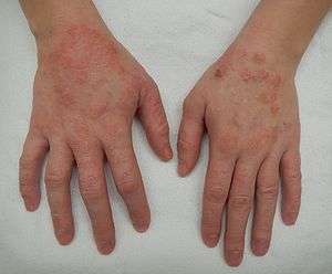
| A moderate case of dermatitis of the hands | |
| Specialty | Dermatology |
| Symptoms | Itchiness, red skin, rash[1] |
| Complications | Skin infection[2] |
| Usual onset | Childhood[1][2] |
| Causes | Atopic dermatitis, allergic contact dermatitis, irritant contact dermatitis, stasis dermatitis[1][2] |
| Diagnostic method | Based on symptom[1] |
| Differential diagnosis | Scabies, psoriasis, dermatitis herpetiformis, lichen simplex chronicus[3] |
| Treatment | Moisturizers, steroid creams, antihistamines[2][4] |
| Frequency | 245 million in 2015[5] (3.34% of world population) |
Dermatitis includes atopic dermatitis, allergic contact dermatitis, irritant contact dermatitis and stasis dermatitis.[1][2] The exact cause of the condition is often unclear.[2] Cases may involve a combination of allergy and poor venous return.[1] The type of dermatitis is generally determined by the person's history and the location of the rash.[1] For example, irritant dermatitis often occurs on the hands of those who frequently get them wet.[1] Allergic contact dermatitis occurs upon exposure to an allergen, causing a hypersensitivity reaction in the skin.[1]
Treatment of atopic dermatitis is typically with moisturizers and steroid creams.[4] The steroid creams should generally be of mid- to high strength and used for less than two weeks at a time, as side effects can occur.[6] Antibiotics may be required if there are signs of skin infection.[2] Contact dermatitis is typically treated by avoiding the allergen or irritant.[7][8] Antihistamines may help with sleep and decrease nighttime scratching.[2]
Dermatitis was estimated to affect 245 million people globally in 2015,[5] or 3.34% of the world population. Atopic dermatitis is the most common type and generally starts in childhood.[1][2] In the United States, it affects about 10–30% of people.[2] Contact dermatitis is twice as common in females than males.[9] Allergic contact dermatitis affects about 7% of people at some point in their lives.[10] Irritant contact dermatitis is common, especially among people with certain occupations; exact rates are unclear.[11]
Signs and symptoms

Dermatitis symptoms vary with all different forms of the condition. They range from skin rashes to bumpy rashes or including blisters.[12] Although every type of dermatitis has different symptoms, there are certain signs that are common for all of them, including redness of the skin, swelling, itching and skin lesions with sometimes oozing and scarring. Also, the area of the skin on which the symptoms appear tends to be different with every type of dermatitis, whether on the neck, wrist, forearm, thigh or ankle. Although the location may vary, the primary symptom of this condition is itchy skin. More rarely, it may appear on the genital area, such as the vulva or scrotum.[13][14] Symptoms of this type of dermatitis may be very intense and may come and go. Irritant contact dermatitis is usually more painful than itchy.
Although the symptoms of atopic dermatitis vary from person to person, the most common symptoms are dry, itchy, red skin. Typical affected skin areas include the folds of the arms, the back of the knees, wrists, face and hands. Perioral dermatitis refers to a red bumpy rash around the mouth.[15]
Dermatitis herpetiformis symptoms include itching, stinging and a burning sensation. Papules and vesicles are commonly present.[16] The small red bumps experienced in this type of dermatitis are usually about 1 cm in size, red in color and may be found symmetrically grouped or distributed on the upper or lower back, buttocks, elbows, knees, neck, shoulders, and scalp. Less frequently, the rash may appear inside the mouth or near the hairline.
The symptoms of seborrheic dermatitis, on the other hand, tend to appear gradually, from dry or greasy scaling of the scalp (dandruff) to scaling of facial areas, sometimes with itching, but without hair loss.[17] In newborns, the condition causes a thick and yellowish scalp rash, often accompanied by a diaper rash. In severe cases, symptoms may appear along the hairline, behind the ears, on the eyebrows, on the bridge of the nose, around the nose, on the chest, and on the upper back.[18]
 Dermatitis
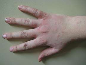
 More severe dermatitis
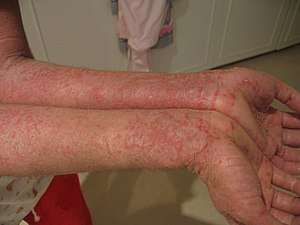
 A patch of dermatitis that has been scratched
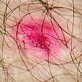
 Complex dermatitis
Cause
The cause of dermatitis is unknown but is presumed to be a combination of genetic and environmental factors.[2]
Environmental
The hygiene hypothesis postulates that the cause of asthma, eczema, and other allergic diseases is an unusually clean environment in childhood which leads to an insufficient human microbiota. It is supported by epidemiologic studies for asthma.[19] The hypothesis states that exposure to bacteria and other immune system modulators is important during development, and missing out on this exposure increases the risk for asthma and allergy.
While it has been suggested that eczema may sometimes be an allergic reaction to the excrement from house dust mites,[20] with up to 5% of people showing antibodies to the mites,[21] the overall role this plays awaits further corroboration.[22]
Genetic
A number of genes have been associated with eczema, one of which is filaggrin.[4] Genome-wide studies found three new genetic variants associated with eczema: OVOL1, ACTL9 and IL4-KIF3A.[23]
Eczema occurs about three times more frequently in individuals with celiac disease and about two times more frequently in relatives of those with celiac disease, potentially indicating a genetic link between the conditions.[24][25]
Pathophysiology
Eczema can be characterized by spongiosis which allows inflammatory mediators to accumulate. Different dendritic cells sub types, such as Langerhans cells, inflammatory dendritic epidermal cells and plasmacytoid dendritic cells have a role to play.[26][27]
Diagnosis
Diagnosis of eczema is based mostly on the history and physical examination.[4] In uncertain cases, skin biopsy may be taken for a histopathologic diagnosis of dermatitis.[28] Those with eczema may be especially prone to misdiagnosis of food allergies.[29]
Patch tests are used in the diagnosis of allergic contact dermatitis.[30][31]
Classification
The term "eczema" refers to a set of clinical characteristics. Classification of the underlying diseases has been haphazard with numerous different classification systems, and many synonyms being used to describe the same condition.
A type of dermatitis may be described by location (e.g., hand eczema), by specific appearance (eczema craquele or discoid) or by possible cause (varicose eczema). Further adding to the confusion, many sources use the term eczema interchangeably for the most common type: atopic dermatitis.
The European Academy of Allergology and Clinical Immunology (EAACI) published a position paper in 2001, which simplifies the nomenclature of allergy-related diseases, including atopic and allergic contact eczemas.[32] Non-allergic eczemas are not affected by this proposal.
Histopathologic classification
By histopathology, superficial dermatitis (in the epidermis, papillary dermis, and superficial vascular plexus) can basically be classified into either of the following groups:[33]
- Vesiculobullous lesions
- Pustular dermatosis
- Non vesicullobullous, non-pustular
- With epidermal changes
- Without epidermal changes. These characteristically have a superficial perivascular inflammatory infiltrate, and can be classified by type of cell infiltrate:[33]
- Lymphocytic (most common)
- Lymphoeosinophilic
- Lymphoplasmacytic
- Mast cell
- Lymphohistiocytic
- Neutrophilic
Terminology
There are several types of dermatitis including atopic dermatitis, contact dermatitis, stasis dermatitis and seborrheic eczema.[2] Many use the term dermatitis and eczema synonymously.[1]
Others use the term eczema to specifically mean atopic dermatitis.[34][35][36] Atopic dermatitis is also known as atopic eczema.[4] In some languages, dermatitis and eczema mean the same thing, while in other languages dermatitis implies an acute condition and eczema a chronic one.[37]
Common types
Diagnosis of types may be indicated by codes defined according to International Statistical Classification of Diseases and Related Health Problems (ICD).
Atopic
Atopic dermatitis is an allergic disease believed to have a hereditary component and often runs in families whose members have asthma. Itchy rash is particularly noticeable on head and scalp, neck, inside of elbows, behind knees, and buttocks. It is very common in developed countries and rising. Irritant contact dermatitis is sometimes misdiagnosed as atopic dermatitis. Stress can cause atopic dermatitis to worsen.[38]
Contact
Contact dermatitis is of two types: allergic (resulting from a delayed reaction to an allergen, such as poison ivy, nickel, or Balsam of Peru),[39] and irritant (resulting from direct reaction to a detergent, such as sodium lauryl sulfate, for example).
Some substances act both as allergen and irritant (wet cement, for example). Other substances cause a problem after sunlight exposure, bringing on phototoxic dermatitis. About three quarters of cases of contact eczema are of the irritant type, which is the most common occupational skin disease. Contact eczema is curable, provided the offending substance can be avoided and its traces removed from one's environment. (ICD-10 L23; L24; L56.1; L56.0)
Seborrhoeic
Seborrhoeic dermatitis or seborrheic dermatitis ("cradle cap" in infants) is a condition sometimes classified as a form of eczema that is closely related to dandruff. It causes dry or greasy peeling of the scalp, eyebrows, and face, and sometimes trunk. In newborns, it causes a thick, yellow, crusty scalp rash called cradle cap, which seems related to lack of biotin and is often curable. (ICD-10 L21; L21.0)
Less common types
Dyshidrosis
Dyshidrosis (dyshidrotic eczema, pompholyx, vesicular palmoplantar dermatitis) only occurs on palms, soles, and sides of fingers and toes. Tiny opaque bumps called vesicles, thickening, and cracks are accompanied by itching, which gets worse at night. A common type of hand eczema, it worsens in warm weather. (ICD-10 L30.1)
Discoid
Discoid eczema (nummular eczema, exudative eczema, microbial eczema) is characterized by round spots of oozing or dry rash, with clear boundaries, often on lower legs. It is usually worse in winter. Cause is unknown, and the condition tends to come and go. (ICD-10 L30.0)
Venous
Venous eczema (gravitational eczema, stasis dermatitis, varicose eczema) occurs in people with impaired circulation, varicose veins, and edema, and is particularly common in the ankle area of people over 50. There is redness, scaling, darkening of the skin, and itching. The disorder predisposes to leg ulcers. (ICD-10 I83.1)
Herpetiformis
Dermatitis herpetiformis (Duhring's disease) causes an intensely itchy and typically symmetrical rash on arms, thighs, knees, and back. It is directly related to celiac disease, can often be put into remission with an appropriate diet, and tends to get worse at night. (ICD-10 L13.0)
Neurodermatitis
Neurodermatitis (lichen simplex chronicus, localized scratch dermatitis) is an itchy area of thickened, pigmented eczema patch that results from habitual rubbing and scratching. Usually, there is only one spot. Often curable through behaviour modification and anti-inflammatory medication. Prurigo nodularis is a related disorder showing multiple lumps. (ICD-10 L28.0; L28.1)
Autoeczematization
Autoeczematization (id reaction, auto sensitization) is an eczematous reaction to an infection with parasites, fungi, bacteria, or viruses. It is completely curable with the clearance of the original infection that caused it. The appearance varies depending on the cause. It always occurs some distance away from the original infection. (ICD-10 L30.2)
Viral
There are eczemas overlaid by viral infections (eczema herpeticum or vaccinatum), and eczemas resulting from underlying disease (e.g., lymphoma). Eczemas originating from ingestion of medications, foods, and chemicals, have not yet been clearly systematized. Other rare eczematous disorders exist in addition to those listed here.
Prevention
Exclusive breastfeeding of infants during at least the first few months may decrease the risk.[40] There is no good evidence that a mother's diet during pregnancy or breastfeeding affects the risk,[40] nor is there evidence that delayed introduction of certain foods is useful.[40] There is tentative evidence that probiotics in infancy may reduce rates but it is insufficient to recommend its use.[41]
Certain military and healthcare personnel who might come into contact with the smallpox are still vaccinated against the virus.[42] Those who also have eczema should not receive the smallpox vaccination due to risk of developing eczema vaccinatum, a potentially severe and sometimes fatal complication.[43]
Management
There is no known cure for some types of dermatitis, with treatment aiming to control symptoms by reducing inflammation and relieving itching. Contact dermatitis is treated by avoiding what is causing it.
Lifestyle
Bathing once or more a day is recommended, usually for five to ten minutes in warm water.[4][44] Soaps should be avoided as they tend to strip the skin of natural oils and lead to excessive dryness.[45] The American Academy of Dermatology suggests using a controlled amount of bleach diluted in a bath to help with atopic dermatitis.[46]
There has not been adequate evaluation of changing the diet to reduce eczema.[47][48] There is some evidence that infants with an established egg allergy may have a reduction in symptoms if eggs are eliminated from their diets.[47] Benefits have not been shown for other elimination diets, though the studies are small and poorly executed.[47][48] Establishing that there is a food allergy before dietary change could avoid unnecessary lifestyle changes.[47]
People can wear clothing designed to manage the itching, scratching and peeling.[49]
House dust mite reduction and avoidance measures have been studied in low quality trials and have not shown evidence of improving eczema.[50]
Moisturizers
Low-quality evidence indicates that moisturizing agents (emollients) may reduce eczema severity and lead to fewer flares.[51] In children, oil–based formulations appear to be better and water–based formulations are not recommended.[4] It is unclear if moisturizers that contain ceramides are more or less effective than others.[52] Products that contain dyes, perfumes, or peanuts should not be used.[4] Occlusive dressings at night may be useful.[4]
Some moisturizers or barrier creams may reduce irritation in occupational irritant hand dermatitis,[53] a skin disease that can affect people in jobs that regularly come into contact with water, detergents, chemicals or other irritants.[53] Some emollients may reduce the number of flares in people with dermatitis.[54]
Medications
Corticosteroids
If symptoms are well controlled with moisturizers, steroids may only be required when flares occur.[4] Corticosteroids are effective in controlling and suppressing symptoms in most cases.[55] Once daily use is generally enough.[4] For mild-moderate eczema a weak steroid may be used (e.g., hydrocortisone), while in more severe cases a higher-potency steroid (e.g., clobetasol propionate) may be used. In severe cases, oral or injectable corticosteroids may be used. While these usually bring about rapid improvements, they have greater side effects.
Long term use of topical steroids may result in skin atrophy, stria, telangiectasia.[4] Their use on delicate skin (face or groin) is therefore typically with caution.[4] They are, however, generally well tolerated.[56] Red burning skin, where the skin turns red upon stopping steroid use, has been reported among adults who use topical steroids at least daily for more than a year.[57]
Antihistamines
There is little evidence supporting the efficacy of antihistamine for the relief of dermatitis.[4][58] Sedative antihistamines, such as diphenhydramine, may be useful in those who are unable to sleep due to eczema.[4] Second generation antihistamines have minimal evidence of benefit.[59] Of the second generation antihistamines studied, fexofenadine is the only one to show evidence of improvement in itching with minimal side effects.[59]
Immunosuppressants
Topical immunosuppressants like pimecrolimus and tacrolimus may be better in the short term and appear equal to steroids after a year of use.[60] Their use is reasonable in those who do not respond to or are not tolerant of steroids.[61][62] Treatments are typically recommended for short or fixed periods of time rather than indefinitely.[4][63] Tacrolimus 0.1% has generally proved more effective than pimecrolimus, and equal in effect to mid-potency topical steroids.[64] There is no link to increased risk of cancer from topical use of 1% pimecrolimus cream.[63]
When eczema is severe and does not respond to other forms of treatment, systemic immunosuppressants are sometimes used. Immunosuppressants can cause significant side effects and some require regular blood tests. The most commonly used are ciclosporin, azathioprine, and methotrexate.
Light therapy
Light therapy using ultraviolet light has tentative support but the quality of the evidence is not very good.[65] A number of different types of light may be used including UVA and UVB;[66] in some forms of treatment, light sensitive chemicals such as psoralen are also used. Overexposure to ultraviolet light carries its own risks, particularly that of skin cancer.[67]
Alternative medicine
Limited evidence suggests that acupuncture may reduce itching in those affected by atopic dermatitis.[68] There is currently no scientific evidence for the claim that sulfur treatment relieves eczema.[69] It is unclear whether Chinese herbs help or harm.[70] Dietary supplements are commonly used by people with eczema.[71] Neither evening primrose oil nor borage seed oil taken orally have been shown to be effective.[72] Both are associated with gastrointestinal upset.[72] Probiotics are likely to make little to no difference in symptoms.[73] There is insufficient evidence to support the use of zinc, selenium, vitamin D, vitamin E, pyridoxine (vitamin B6), sea buckthorn oil, hempseed oil, sunflower oil, or fish oil as dietary supplements.[71]
Chiropractic spinal manipulation lacks evidence to support its use for dermatitis.[74] There is little evidence supporting the use of psychological treatments.[75] While dilute bleach baths have been used for infected dermatitis there is little evidence for this practice.[76]
Prognosis
Most cases are well managed with topical treatments and ultraviolet light.[4] About 2% of cases are not.[4] In more than 60% of young children, the condition subsides by adolescence.[4]
Epidemiology
Globally dermatitis affected approximately 230 million people as of 2010 (3.5% of the population).[77] Dermatitis is most commonly seen in infancy, with female predominance of eczema presentations occurring during the reproductive period of 15–49 years.[78] In the UK about 20% of children have the condition, while in the United States about 10% are affected.[4]
Although little data on the rates of eczema over time exists prior to the 1940s, the rate of eczema has been found to have increased substantially in the latter half of the 20th century, with eczema in school-aged children being found to increase between the late 1940s and 2000.[79] In the developed world there has been rise in the rate of eczema over time. The incidence and lifetime prevalence of eczema in England has been seen to increase in recent times.[4][80]
Dermatitis affected about 10% of U.S. workers in 2010, representing over 15 million workers with dermatitis. Prevalence rates were higher among females than among males, and among those with some college education or a college degree compared to those with a high school diploma or less. Workers employed in healthcare and social assistance industries and life, physical, and social science occupations had the highest rates of reported dermatitis. About 6% of dermatitis cases among U.S. workers were attributed to work by a healthcare professional, indicating that the prevalence rate of work-related dermatitis among workers was at least 0.6%.[81]
History
from ἐκζέ-ειν ekzé-ein,
from ἐκ ek "out" + ζέ-ειν zé-ein "to boil"
(OED)
The term "atopic dermatitis" was coined in 1933 by Wise and Sulzberger.[83] Sulfur as a topical treatment for eczema was fashionable in the Victorian and Edwardian eras.[69]
The word dermatitis is from the Greek δέρμα derma "skin" and -ῖτις -itis "inflammation" and eczema is from Greek: ἔκζεμα ekzema "eruption".[84]
Society and culture
The terms "hypoallergenic" and "doctor tested" are not regulated,[85] and no research has been done showing that products labeled "hypoallergenic" are less problematic than any others.
Research
Monoclonal antibodies are under preliminary research to determine their potential as treatments for atopic dermatitis, with only dupilumab showing evidence of efficacy, as of 2018.[86][87]
References
- Nedorost, Susan T. (2012). Generalized Dermatitis in Clinical Practice. Springer Science & Business Media. pp. 1–3, 9, 13–14. ISBN 9781447128977. Archived from the original on 15 August 2016. Retrieved 29 July 2016.
- "Handout on Health: Atopic Dermatitis (A type of eczema)". NIAMS. May 2013. Archived from the original on 30 May 2015. Retrieved 29 July 2016.
- Ferri, Fred F. (2010). Ferri's differential diagnosis : a practical guide to the differential diagnosis of symptoms, signs, and clinical disorders (2nd ed.). Philadelphia, PA: Elsevier/Mosby. p. Chapter D. ISBN 978-0323076999.
- McAleer MA, Flohr C, Irvine AD (July 2012). "Management of difficult and severe eczema in childhood" (PDF). BMJ. 345: e4770. doi:10.1136/bmj.e4770. hdl:2262/75991. PMID 22826585.
- Vos T, Allen C, Arora M, Barber RM, Bhutta ZA, Brown A, et al. (GBD 2015 Disease and Injury Incidence and Prevalence Collaborators) (October 2016). "Global, regional, and national incidence, prevalence, and years lived with disability for 310 diseases and injuries, 1990-2015: a systematic analysis for the Global Burden of Disease Study 2015". Lancet. 388 (10053): 1545–1602. doi:10.1016/S0140-6736(16)31678-6. PMC 5055577. PMID 27733282.
- Habif (2015). Clinical Dermatology (6 ed.). Elsevier Health Sciences. p. 171. ISBN 9780323266079. Archived from the original on 17 August 2016. Retrieved 5 July 2016.
- Mowad CM, Anderson B, Scheinman P, Pootongkam S, Nedorost S, Brod B (June 2016). "Allergic contact dermatitis: Patient management and education". Journal of the American Academy of Dermatology. 74 (6): 1043–54. doi:10.1016/j.jaad.2015.02.1144. PMID 27185422.
- Lurati AR (February 2015). "Occupational risk assessment and irritant contact dermatitis". Workplace Health & Safety. 63 (2): 81–7, quiz 88. doi:10.1177/2165079914565351. PMID 25881659. S2CID 40077567.
- Adkinson, N. Franklin (2014). Middleton's allergy : principles and practice (8 ed.). Philadelphia: Elsevier Saunders. p. 566. ISBN 9780323085939. Archived from the original on 15 August 2016.
- "128.4". Rook's Textbook of Dermatology, 4 Volume Set (9 ed.). John Wiley & Sons. 2016. ISBN 9781118441176. Archived from the original on 15 August 2016. Retrieved 29 July 2016.
- Frosch, Peter J. (2013). Textbook of Contact Dermatitis (2 ed.). Berlin, Heidelberg: Springer Berlin Heidelberg. p. 42. ISBN 9783662031049. Archived from the original on 16 August 2016.
- Caproni M, Antiga E, Melani L, Fabbri P (June 2009). "Guidelines for the diagnosis and treatment of dermatitis herpetiformis". Journal of the European Academy of Dermatology and Venereology. 23 (6): 633–8. doi:10.1111/j.1468-3083.2009.03188.x. PMID 19470076.
- "Neurodermatitis (lichen simplex)". DermNet New Zealand Trust. 2017. Archived from the original on 2 February 2017. Retrieved 29 January 2017.
- "Neurodermatitis". Mayo Clinic. 2015. Archived from the original on 16 June 2010. Retrieved 6 November 2010.
- "Periorificial dermatitis". DermNet New Zealand Trust. 2017. Archived from the original on 2 February 2017. Retrieved 29 January 2017.
- "Dermatitis herpetiformis". DermNet New Zealand Trust. 2017. Archived from the original on 2 February 2017. Retrieved 29 January 2017.
- "Seborrheic dermatitis". DermNet New Zealand Trust. 2017. Archived from the original on 2 February 2017. Retrieved 29 January 2017.
- "Seborrheic Dermatitis". Merck Manual, Consumer Version.
- Bufford JD, Gern JE (May 2005). "The hygiene hypothesis revisited". Immunology and Allergy Clinics of North America. 25 (2): 247–62, v–vi. doi:10.1016/j.iac.2005.03.005. PMID 15878454.
- Carswell F, Thompson S (July 1986). "Does natural sensitisation in eczema occur through the skin?". Lancet. 2 (8497): 13–5. doi:10.1016/S0140-6736(86)92560-2. PMID 2873316.
- Henszel Ł, Kuźna-Grygiel W (2006). "[House dust mites in the etiology of allergic diseases]". Annales Academiae Medicae Stetinensis (in Polish). 52 (2): 123–7. PMID 17633128.
- Atopic Dermatitis at eMedicine
- Paternoster L, Standl M, Chen CM, Ramasamy A, Bønnelykke K, Duijts L, et al. (December 2011). "Meta-analysis of genome-wide association studies identifies three new risk loci for atopic dermatitis". Nature Genetics. 44 (2): 187–92. doi:10.1038/ng.1017. PMC 3272375. PMID 22197932.
- Caproni M, Bonciolini V, D'Errico A, Antiga E, Fabbri P (2012). "Celiac disease and dermatologic manifestations: many skin clue to unfold gluten-sensitive enteropathy". Gastroenterology Research and Practice. Hindawi Publishing Corporation. 2012: 952753. doi:10.1155/2012/952753. PMC 3369470. PMID 22693492.
- Ciacci C, Cavallaro R, Iovino P, Sabbatini F, Palumbo A, Amoruso D, et al. (June 2004). "Allergy prevalence in adult celiac disease". The Journal of Allergy and Clinical Immunology. 113 (6): 1199–203. doi:10.1016/j.jaci.2004.03.012. PMID 15208605.
- Allam JP, Novak N (January 2006). "The pathophysiology of atopic eczema". Clinical and Experimental Dermatology. 31 (1): 89–93. doi:10.1111/j.1365-2230.2005.01980.x. PMID 16309494.
- Ulf D, Eyerich K, Ring J (October 2007). "Eczema Pathophysiology - World Allergy Organization". www.worldallergy.org. Archived from the original on 2 February 2017. Retrieved 28 January 2017. Cite journal requires
|journal=(help) - "Eczema". University of Maryland Medical Center. Archived from the original on 27 July 2016.
- Atkins D (March 2008). "Food allergy: diagnosis and management". Primary Care. 35 (1): 119–40, vii. doi:10.1016/j.pop.2007.09.003. PMID 18206721.
- Johansen JD, Frosch PJ, Lepoittevin J (29 September 2010). Contact Dermatitis. ISBN 9783642038273. Archived from the original on 5 July 2014. Retrieved 21 April 2014.
- Fisher AA (2008). Fisher's Contact Dermatitis. ISBN 9781550093780. Retrieved 21 April 2014.
- Johansson SG, Hourihane JO, Bousquet J, Bruijnzeel-Koomen C, Dreborg S, Haahtela T, et al. (September 2001). "A revised nomenclature for allergy. An EAACI position statement from the EAACI nomenclature task force". Allergy. 56 (9): 813–24. doi:10.1034/j.1398-9995.2001.t01-1-00001.x. PMID 11551246.
- Alsaad KO, Ghazarian D (December 2005). "My approach to superficial inflammatory dermatoses". Journal of Clinical Pathology. 58 (12): 1233–41. doi:10.1136/jcp.2005.027151. PMC 1770784. PMID 16311340.
- "Eczema". ACP medicine. Archived from the original on 10 January 2014. Retrieved 9 January 2014.
- Bershad SV (November 2011). "In the clinic. Atopic dermatitis (eczema)". Annals of Internal Medicine. 155 (9): ITC51–15, quiz ITC516. doi:10.7326/0003-4819-155-9-201111010-01005. PMID 22041966.
- ICD 10: Diseases of the skin and subcutaneous tissue (L00-L99) – Dermatitis and eczema (L20-L30) Archived 9 January 2014 at the Wayback Machine
- Ring J, Przybilla B, Ruzicka T (2006). Handbook of atopic eczema. Birkhäuser. p. 4. ISBN 978-3-540-23133-2. Retrieved 4 May 2010.
- Atopic Dermatitis National Eczema Association.
- "Balsam of Peru contact allergy". Dermnetnz.org. 28 December 2013. Archived from the original on 5 March 2014. Retrieved 5 March 2014.
- Greer FR, Sicherer SH, Burks AW (April 2019). "The Effects of Early Nutritional Interventions on the Development of Atopic Disease in Infants and Children: The Role of Maternal Dietary Restriction, Breastfeeding, Hydrolyzed Formulas, and Timing of Introduction of Allergenic Complementary Foods". Pediatrics. 143 (4): e20190281. doi:10.1542/peds.2019-0281. PMID 30886111.
- Kalliomäki M, Antoine JM, Herz U, Rijkers GT, Wells JM, Mercenier A (March 2010). "Guidance for substantiating the evidence for beneficial effects of probiotics: prevention and management of allergic diseases by probiotics". The Journal of Nutrition. 140 (3): 713S–21S. doi:10.3945/jn.109.113761. PMID 20130079.
- "DoD Details Military Smallpox Vaccination Program". www.navy.mil. Retrieved 10 January 2019.
- "CDC Smallpox | Smallpox (Vaccinia) Vaccine Contraindications (Info for Clinicians)". Emergency.cdc.gov. 7 February 2007. Archived from the original on 25 January 2010. Retrieved 7 February 2010.
- "Coping with atopic dermatitis". 2017. Retrieved 11 September 2017.
- Gutman AB, Kligman AM, Sciacca J, James WD (December 2005). "Soak and smear: a standard technique revisited". Archives of Dermatology. 141 (12): 1556–9. doi:10.1001/archderm.141.12.1556. PMID 16365257.
- "Atopic dermatitis: Bleach bath therapy". www.aad.org. Retrieved 4 May 2020.
- Bath-Hextall F, Delamere FM, Williams HC (January 2008). Bath-Hextall FJ (ed.). "Dietary exclusions for established atopic eczema". The Cochrane Database of Systematic Reviews (1): CD005203. doi:10.1002/14651858.CD005203.pub2. PMC 6885041. PMID 18254073. Archived from the original on 21 October 2013.
- Institute for Quality and Efficiency in Health Care. "Eczema: Can eliminating particular foods help?". Informed Health Online. Institute for Quality and Efficiency in Health Care. Archived from the original on 21 October 2013. Retrieved 24 June 2013.
- Ricci G, Patrizi A, Bellini F, Medri M (2006). Use of textiles in atopic dermatitis: care of atopic dermatitis. Current Problems in Dermatology. 33. pp. 127–43. doi:10.1159/000093940. ISBN 978-3-8055-8121-9. PMID 16766885.
- Nankervis H, Pynn EV, Boyle RJ, Rushton L, Williams HC, Hewson DM, Platts-Mills T (January 2015). "House dust mite reduction and avoidance measures for treating eczema". The Cochrane Database of Systematic Reviews. 1: CD008426. doi:10.1002/14651858.CD008426.pub2. hdl:10044/1/21547. PMID 25598014.
- van Zuuren EJ, Fedorowicz Z, Christensen R, Lavrijsen A, Arents BW (February 2017). "Emollients and moisturisers for eczema". The Cochrane Database of Systematic Reviews. 2: CD012119. doi:10.1002/14651858.CD012119.pub2. PMC 6464068. PMID 28166390.
- Jungersted JM, Agner T (August 2013). "Eczema and ceramides: an update". Contact Dermatitis. 69 (2): 65–71. doi:10.1111/cod.12073. PMID 23869725.
- Bauer A, Rönsch H, Elsner P, Dittmar D, Bennett C, Schuttelaar ML, et al. (April 2018). "Interventions for preventing occupational irritant hand dermatitis" (PDF). The Cochrane Database of Systematic Reviews. 4: CD004414. doi:10.1002/14651858.CD004414.pub3. PMC 6494486. PMID 29708265. Archived from the original (PDF) on 6 March 2020. Retrieved 26 June 2019.
- van Zuuren EJ, Fedorowicz Z, Christensen R, Lavrijsen A, Arents BW (February 2017). "Emollients and moisturisers for eczema". The Cochrane Database of Systematic Reviews. 2: CD012119. doi:10.1002/14651858.CD012119.pub2. PMC 6464068. PMID 28166390.
- Hoare C, Li Wan Po A, Williams H (2000). "Systematic review of treatments for atopic eczema". Health Technology Assessment. 4 (37): 1–191. doi:10.3310/hta4370. PMC 4782813. PMID 11134919. Archived from the original on 7 February 2009. Retrieved 18 November 2009.
- Bewley A (May 2008). "Expert consensus: time for a change in the way we advise our patients to use topical corticosteroids". The British Journal of Dermatology. 158 (5): 917–20. doi:10.1111/j.1365-2133.2008.08479.x. PMID 18294314.
- Oakley, M.D., Amanda. "Topical corticosteroid withdrawal". DermNet NZ. DermNet New Zealand Trust. Archived from the original on 16 March 2016.
- Apfelbacher CJ, van Zuuren EJ, Fedorowicz Z, Jupiter A, Matterne U, Weisshaar E (February 2013). "Oral H1 antihistamines as monotherapy for eczema". The Cochrane Database of Systematic Reviews (2): CD007770. doi:10.1002/14651858.CD007770.pub2. PMC 6823266. PMID 23450580.
- Matterne U, Böhmer MM, Weisshaar E, Jupiter A, Carter B, Apfelbacher CJ (January 2019). "Oral H1 antihistamines as 'add-on' therapy to topical treatment for eczema". The Cochrane Database of Systematic Reviews. 1: CD012167. doi:10.1002/14651858.CD012167.pub2. PMC 6360926. PMID 30666626.
- Shams K, Grindlay DJ, Williams HC (August 2011). "What's new in atopic eczema? An analysis of systematic reviews published in 2009-2010". Clinical and Experimental Dermatology. 36 (6): 573–7, quiz 577–8. doi:10.1111/j.1365-2230.2011.04078.x. PMID 21718344.
- Carr WW (August 2013). "Topical calcineurin inhibitors for atopic dermatitis: review and treatment recommendations". Paediatric Drugs. 15 (4): 303–10. doi:10.1007/s40272-013-0013-9. PMC 3715696. PMID 23549982.
- "Atopic eczema - Treatment". NHS Choices, London, UK. 12 February 2016. Archived from the original on 16 January 2017. Retrieved 27 January 2017.
- "Medication Guide. Elidel® (pimecrolimus) Cream, 1%" (PDF). US Food and Drug Administration. March 2014. Archived (PDF) from the original on 11 February 2017. Retrieved 27 January 2017.
- Torley D, Futamura M, Williams HC, Thomas KS (July 2013). "What's new in atopic eczema? An analysis of systematic reviews published in 2010-11". Clinical and Experimental Dermatology. 38 (5): 449–56. doi:10.1111/ced.12143. PMID 23750610.
- Gambichler T (March 2009). "Management of atopic dermatitis using photo(chemo)therapy". Archives of Dermatological Research. 301 (3): 197–203. doi:10.1007/s00403-008-0923-5. PMID 19142651.
- Meduri NB, Vandergriff T, Rasmussen H, Jacobe H (August 2007). "Phototherapy in the management of atopic dermatitis: a systematic review". Photodermatology, Photoimmunology & Photomedicine. 23 (4): 106–12. doi:10.1111/j.1600-0781.2007.00291.x. PMID 17598862.
- Stöppler MC (31 May 2007). "Psoriasis PUVA Treatment Can Increase Melanoma Risk". MedicineNet. Archived from the original on 29 September 2007. Retrieved 17 October 2007.
- Vieira BL, Lim NR, Lohman ME, Lio PA (December 2016). "Complementary and Alternative Medicine for Atopic Dermatitis: An Evidence-Based Review". American Journal of Clinical Dermatology (Review). 17 (6): 557–581. doi:10.1007/s40257-016-0209-1. PMID 27388911. S2CID 35460522.
- "Sulfur". University of Maryland Medical Center. 1 April 2002. Archived from the original on 5 August 2012. Retrieved 15 October 2007.
- Armstrong NC, Ernst E (August 1999). "The treatment of eczema with Chinese herbs: a systematic review of randomized clinical trials". British Journal of Clinical Pharmacology. 48 (2): 262–4. doi:10.1046/j.1365-2125.1999.00004.x. PMC 2014284. PMID 10417508.
- Bath-Hextall FJ, Jenkinson C, Humphreys R, Williams HC (February 2012). Bath-Hextall FJ (ed.). "Dietary supplements for established atopic eczema". The Cochrane Database of Systematic Reviews. 2 (2): CD005205. doi:10.1002/14651858.CD005205.pub3. PMC 6517242. PMID 22336810.
- Bamford JT, Ray S, Musekiwa A, van Gool C, Humphreys R, Ernst E (April 2013). Bamford JT (ed.). "Oral evening primrose oil and borage oil for eczema". The Cochrane Database of Systematic Reviews. 4 (4): CD004416. doi:10.1002/14651858.CD004416.pub2. PMID 23633319.
- Makrgeorgou A, Leonardi-Bee J, Bath-Hextall FJ, Murrell DF, Tang ML, Roberts A, Boyle RJ (November 2018). "Probiotics for treating eczema". The Cochrane Database of Systematic Reviews. 11: CD006135. doi:10.1002/14651858.CD006135.pub3. PMC 6517242. PMID 30480774.
- Eldred DC, Tuchin PJ (November 1999). "Treatment of acute atopic eczema by chiropractic care. A case study". Australasian Chiropractic & Osteopathy. 8 (3): 96–101. PMC 2051093. PMID 17987197.
- Ersser SJ, Cowdell F, Latter S, Gardiner E, Flohr C, Thompson AR, et al. (January 2014). "Psychological and educational interventions for atopic eczema in children". The Cochrane Database of Systematic Reviews (1): CD004054. doi:10.1002/14651858.CD004054.pub3. PMC 6457897. PMID 24399641.
- Barnes TM, Greive KA (November 2013). "Use of bleach baths for the treatment of infected atopic eczema". The Australasian Journal of Dermatology. 54 (4): 251–8. doi:10.1111/ajd.12015. PMID 23330843.
- Vos T, Flaxman AD, Naghavi M, Lozano R, Michaud C, Ezzati M, et al. (December 2012). "Years lived with disability (YLDs) for 1160 sequelae of 289 diseases and injuries 1990-2010: a systematic analysis for the Global Burden of Disease Study 2010". Lancet. 380 (9859): 2163–96. doi:10.1016/S0140-6736(12)61729-2. PMC 6350784. PMID 23245607.
- Osman M, Hansell AL, Simpson CR, Hollowell J, Helms PJ (February 2007). "Gender-specific presentations for asthma, allergic rhinitis and eczema in primary care". Primary Care Respiratory Journal. 16 (1): 28–35. doi:10.3132/pcrj.2007.00006. PMC 6634172. PMID 17297524.
- Taylor B, Wadsworth J, Wadsworth M, Peckham C (December 1984). "Changes in the reported prevalence of childhood eczema since the 1939-45 war". Lancet. 2 (8414): 1255–7. doi:10.1016/S0140-6736(84)92805-8. PMID 6150286.
- Simpson CR, Newton J, Hippisley-Cox J, Sheikh A (March 2009). "Trends in the epidemiology and prescribing of medication for eczema in England". Journal of the Royal Society of Medicine. 102 (3): 108–17. doi:10.1258/jrsm.2009.080211. PMC 2746851. PMID 19297652.
- Luckhaupt SE, Dahlhamer JM, Ward BW, Sussell AL, Sweeney MH, Sestito JP, Calvert GM (June 2013). "Prevalence of dermatitis in the working population, United States, 2010 National Health Interview Survey". American Journal of Industrial Medicine. 56 (6): 625–34. doi:10.1002/ajim.22080. PMID 22674651.
- Liddell HG, Scott R. "Ekzema". A Greek-English Lexicon. Tufts University: Perseus.
- Textbook of Atopic Dermatitis. Taylor & Francis. 1 May 2008. p. 1. ISBN 9780203091449. Archived from the original on 28 May 2016.
- "Definition of ECZEMA". www.merriam-webster.com. Archived from the original on 22 February 2016. Retrieved 15 February 2016.
- Murphy LA, White IR, Rastogi SC (May 2004). "Is hypoallergenic a credible term?". Clinical and Experimental Dermatology. 29 (3): 325–7. doi:10.1111/j.1365-2230.2004.01521.x. PMID 15115531.
- Snast I, Reiter O, Hodak E, Friedland R, Mimouni D, Leshem YA (April 2018). "Are Biologics Efficacious in Atopic Dermatitis? A Systematic Review and Meta-Analysis". American Journal of Clinical Dermatology. 19 (2): 145–165. doi:10.1007/s40257-017-0324-7. PMID 29098604. S2CID 4220890.
- Lauffer F, Ring J (2016). "Target-oriented therapy: Emerging drugs for atopic dermatitis". Expert Opinion on Emerging Drugs. 21 (1): 81–9. doi:10.1517/14728214.2016.1146681. PMID 26808004.
External links
| Classification | |
|---|---|
| External resources |
| Look up dermatitis in Wiktionary, the free dictionary. |
| Wikimedia Commons has media related to Dermatitis. |
- Dermatitis at Curlie