Duodenojejunal flexure
The duodenojejunal flexure or duodenojejunal junction is the border between the duodenum and the jejunum.
| Duodenojejunal flexure | |
|---|---|
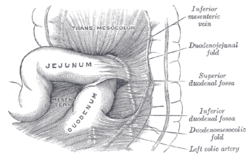
 Superior and inferior duodenal fossæ. | |
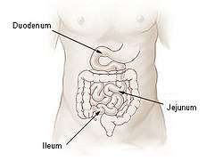
 Small intestine | |
| Details | |
| Identifiers | |
| Latin | flexura duodenojejunalis |
| TA | A05.6.02.009 |
| FMA | 15957 |
| Anatomical terminology | |
Structure
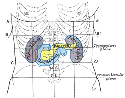
The ascending portion of the duodenum ascends on the left side of the aorta, as far as the level of the upper border of the second lumbar vertebra, where it turns abruptly forward to become the jejunum, forming the duodenojejunal flexure. The duodenojejunal flexure is surrounded by the suspensory muscle of the duodenum.[1] :274
It lies in front of the left Psoas major and left renal vessels, and is covered in front, and partly at the sides, by peritoneum continuous with the left portion of the mesentery.
Clinical relevance
The ligament of Treitz, a peritoneal fold, from the right crus of diaphragm, is an identification point for the duodenojejunal flexure at operation.[2]:85
Additional images
 Duodenojejunal fossa.
See also
References
This article incorporates text in the public domain from page 1170 of the 20th edition of Gray's Anatomy (1918)
- Drake, Richard L.; Vogl, Wayne; Tibbitts, Adam W.M. Mitchell; illustrations by Richard; Richardson, Paul (2005). Gray's anatomy for students. Philadelphia: Elsevier/Churchill Livingstone. ISBN 978-0-8089-2306-0.
- Jacob, S. (2007) Chapter 4: Abdomen; Human anatomy, A clinically-orientated approach.
External links
- Anatomy figure: 37:06-04 at Human Anatomy Online, SUNY Downstate Medical Center - "The large intestine."
- Anatomy photo:39:07-0105 at the SUNY Downstate Medical Center - "Intestines and Pancreas: The Duodenum"
- Anatomy image:8155 at the SUNY Downstate Medical Center