Xanthoma
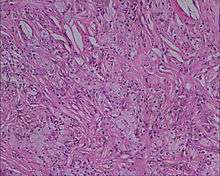
A xanthoma (pl. xanthomas or xanthomata) (condition: xanthomatosis), from Greek ξανθός (xanthós), meaning 'yellow', is a deposition of yellowish cholesterol-rich material that can appear anywhere in the body in various disease states.[2] They are cutaneous manifestations of lipidosis in which lipids accumulate in large foam cells within the skin.[2] They are associated with hyperlipidemias, both primary and secondary types.
| Xanthoma | |
|---|---|
 | |
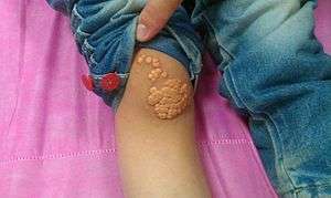
| A patient's knee showing multiple xanthoma tuberosum[1] | |
| Specialty | Gastroenterology, dermatology |
Tendon xanthomas are associated with type II hyperlipidemia, chronic biliary tract obstruction, primary biliary cirrhosis and the rare metabolic disease cerebrotendineous xanthomatosis. Palmar xanthomata and tuberoeruptive xanthomata (over knees and elbows) occur in type III hyperlipidemia.
Types
Xanthelasma

A xanthelasma is a sharply demarcated yellowish collection of cholesterol underneath the skin, usually on or around the eyelids. Strictly, a xanthelasma is a distinct condition, being called a xanthoma only when becoming larger and nodular, assuming tumorous proportions.[3] Still, it is often classified simply as a subtype of xanthoma.[4]
Xanthoma tuberosum
Xanthoma tuberosum (also known as tuberous xanthoma) is characterized by xanthomas located over the joints.[2]:530
Xanthoma tendinosum
Xanthoma tendinosum (also tendon xanthoma or tendinous xanthoma[5]) is clinically characterized by papules and nodules found in the tendons of the hands, feet, and heel.[2]:531 Also associated with familial hypercholesterolemia (FH).[6]
Eruptive xanthoma
Eruptive xanthoma (ILDS E78.220) is clinically characterized by small, yellowish-orange to reddish-brown papules surrounded by an erythematous halo that appear suddenly all over the body, especially the hands, buttocks, and the extensor surfaces of the extremities.[2]:531 It tends to be associated with elevated triglycerides [7]
Xanthoma planum
Xanthoma planum (ILDS D76.370), also known as plane xanthoma, is clinically characterized by bands or rectangular plates (macules) and plaques in the dermis spread diffusely over large areas of the body.[2]:531
Palmar xanthoma
Palmar xanthoma is clinically characterized by yellowish plaques that involve the palms and flexural surfaces of the fingers.[2]:531 Plane xanthomas are characterised by yellowish to orange, flat macules or slightly elevated plaques, often with a central white area which may be localised or generalised. They often arise in the skin folds, especially the palmar creases. They occur in hyperlipoproteinaemia type III and type IIA, and in association with biliary cirrhosis. The presence of palmar xanthomata, like the presence of tendinous xanthomata, is indicative of hypercholesterolaemia.
Tuberoeruptive xanthoma
Tuberoeruptive xanthoma (ILDS E78.210) is clinically characterized by red papules and nodules that appear inflamed and tend to coalesce.[2]:532 Tuberous xanthomata are considered similar, and within the same disease spectrum as eruptive xanthomata.[5]
Other types
Other types of xanthoma identified in the Medical Dictionary include:[8]
- Xanthoma diabeticorum: a type of eruptive xanthoma, often with severe diabetes.
- Xanthoma disseminatum: a rare xanthoma consisting of non-X histiocytes on flexural (folded) surfaces, associated with diabetes insipidus.
- Verrucous xanthoma, or histiocytosis Y: a papilloma of the oral mucosa and skin whereby the connective tissue under the epithelium contains histiocytes.
References
- Kumar et al. Cases Journal 2008
- James, William D.; Berger, Timothy G.; et al. (2006). Andrews' Diseases of the Skin: clinical Dermatology. Saunders Elsevier. ISBN 978-0-7216-2921-6.
- Shields, Carol; Shields, Jerry (2008). Eyelid, conjunctival, and orbital tumors: atlas and textbook. Hagerstwon, MD: Lippincott Williams & Wilkins. ISBN 978-0-7817-7578-6.
- thefreedictionary.com > xanthelasma Citing: The American Heritage Medical Dictionary Copyright 2007, 2004 and Mosby's Medical Dictionary, 8th edition. 2009
- Rapini, Ronald P.; Bolognia, Jean L.; Jorizzo, Joseph L. (2007). Dermatology: 2-Volume Set. St. Louis: Mosby. pp. 1415–16. ISBN 978-1-4160-2999-1.
- van den Bosch, Harrie C.M.; Vos, Louwerens D. (1998). "Achilles'-Tendon Xanthoma in Familial Hypercholesterolemia". New England Journal of Medicine. 338 (22): 1591. doi:10.1056/NEJM199805283382205. ISSN 0028-4793. PMID 9603797.
- Digby M; Belli R; McGraw T; Lee A (2011). "Eruptive xanthomas as a cutaneous manifestation of hypertriglyceridemia: a case report". J Clin Aesthet Dermatol. 4 (1): 44–6. PMC 3030216. PMID 21278899.
- "Xanthoma". Medical Dictionary - Dictionary of Medicine and Human Biology. Retrieved 2015-02-05.
External links
| Classification | |
|---|---|
| External resources |
| Look up xanthoma in Wiktionary, the free dictionary. |

- MedlinePlus Encyclopedia: Xanthoma