Clinical urine tests
Clinical urine tests ( also known as urinalysis, UA) is an examination of urine for certain physical properties, solutes, cells, casts, crystals, organisms, or particulate matter,[1] and mainly serves for medical diagnosis.[2] The word is a portmanteau of the words urine and analysis.[3] Urine culture (a microbiological culture of urine) and urine electrolyte levels are part of urinalysis.
| Urinalysis | |
|---|---|
White blood cells seen under a microscope from a urine sample. | |
| Specialty | clinical pathology |
| MeSH | D016482 |
| Other codes | LOINC Codes for Urinalysis panels |
| MedlinePlus | 003579 |
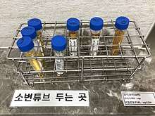
There are three basic components to urinalysis: gross examination, chemical evaluation, and microscopic examination.
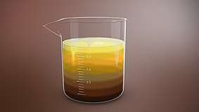
Gross examination targets parameters that can be measured or quantified with the naked eye (or other senses), including volume, color, transparency, odor, and specific gravity.
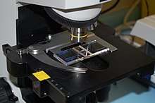
A part of a urinalysis can be performed by using urine test strips, in which the test results can be read as color changes. Another method is light microscopy of urine samples.
Target parameters
Urine test results should always be interpreted using the reference range provided by the laboratory that performed the test, or using information provided by the test strip/device manufacturer.[4][5]
Color

The following are examples of some urine colors and their causes (not a complete listing).
- Nearly colorless: Excessive fluid intake for conditions; untreated diabetes mellitus, diabetes insipidus, and certain types of nephritis.
- Yellow: Distinctly yellow urine may indicate excessive riboflavin (vitamin B2) intake.
- Yellow-amber: Normal.
- Yellow-cloudy: excessive crystals (crystalluria) and/or excessive pus (pyuria).
- Orange: Insufficient fluid intake for conditions; intake of orange substances; intake of phenazopyridine for urinary symptoms.
- Red: Leakage of red blood cells or of hemoglobin from such cells; hemolysis; intake of red substances.
- Dark:
- Reddish-orange: Intake of certain medications or other substances.
- Rusty-yellow to reddish-brown: Intake of certain medications or other substances.
- Dark brown: Intake of certain medications or other substances; damaged muscle (myoglobinuria due to rhabdomyolysis) from extreme exercise or other widespread damage, possibly medication related; altered blood; bilirubinuria; intake of phenolic substances; inadequate porphyrin metabolism; melanin from melanocytic tumors; presence of an abnormal form of hemoglobin, methemoglobin.
- Brownish-black to black: Intake of substances or medications; altered blood; a problem with homogentisic acid metabolism (alkaptonuria), which can also cause dark whites of the eyes and dark-colored internal organs and tissues (ochronosis); Lysol (a product that contains phenols) poisoning; melanin from melanocytic tumors). Paraphenylenediamine is a highly toxic ingredient of hair dye formulations that can cause acute kidney injury and result in black urine.[6]
- Purple due to Purple urine bag syndrome.[6]
- Magenta to purple-red: Presence of phenolphthalein, a stimulant laxative previously found in Ex-Lax.[7]
- Green, or dark with a greenish hue: Jaundice (bilirubinuria); problem with bile metabolism. Recent surgery requiring high doses of propofol infusion.[6] The use of a medication (Uribel) that is similar to phenazopyridine for the relief of urinary symptoms.
- Other colors: Various substances ingested in food or drink, particularly up to 48 hours prior to the presence of colored urine.[8]
Smell
The odor (scent) of urine can normally vary from odorless (when very light colored and dilute) to a much stronger odor when the subject is dehydrated and the urine is concentrated. Brief changes in odor are usually merely interesting and not medically significant. (Example: the abnormal smell many people can detect after eating asparagus.) The urine of diabetics experiencing ketoacidosis (urine containing high levels of ketone bodies) may have a fruity or sweet smell.[9]
Ions and trace minerals
| Target | Lower limit | Upper limit | Unit | Comments | LOINC Codes |
|---|---|---|---|---|---|
| Nitrite | n/a | The presence of nitrites in urine, termed nitrituria, indicates the presence of coliform bacteria. | |||
| Sodium (Na) – per day | 150[11] | 300[11] | mmol / 24 h | A urinalysis is frequently ordered during the workup of acute kidney injury. Full kidney function can be detected through the simple dipstick method. | 2956-1 |
| Potassium (K) – per day | 40[11] | 90[11] | mmol / 24 h | Urine K may be ordered in the workup of hypokalemia. In case of gastrointestinal loss of K, the urine K will be low. In case of renal loss of K, the urine K levels will be high. Decreased levels of urine K are also seen in hypoaldosteronism and adrenal insufficiency. | 2829-0 |
| Urinary calcium (Ca) – per day | 15[12] | 20[12] | mmol / 24 h | An abnormally high level is called hypercalciuria and an abnormally low rate is called hypocalciuria. | 14637-3 |
| 100[12] | 250[12] | mg / 24 hours | 6874-2 | ||
| Phosphate (P) – per day | n/a[11] | 38[11] | mmol / 24 h | Phosphaturia is the hyperexcretion of phosphate in the urine. This condition is divided into primary and secondary types. Primary hyperphosphaturia is characterized by direct excess excretion of phosphate by the kidneys, as from primary kidney dysfunction, and also the direct action of many classes of diuretics on the kidneys. Additionally, secondary causes, including both types of hyperparathyroidism, cause hyperexcretion of phosphate in the urine. | 14881-7 |
A sodium-related parameter is fractional sodium excretion, which is the percentage of the sodium filtered by the kidney which is excreted in the urine. It is a useful parameter in acute kidney failure and oliguria, with a value below 1% indicating a prerenal disease and a value above 3%[13] indicating acute tubular necrosis or other kidney damage.
Proteins and enzymes
| Target | Lower limit | Upper limit | Unit | Comments |
|---|---|---|---|---|
| Protein | 0 | trace amounts[10] / 20 | mg/dl | Proteins may be measured with the Albustix test. Since proteins are very large molecules (macromolecules), they are not normally present in measurable amounts in the glomerular filtrate or in the urine. The detection of protein in urine, called proteinuria, may indicate the permeability of the glomerulus is increased. This may be caused by renal infections or by other diseases that have secondarily affected the kidneys, such as hypertension, diabetes mellitus, jaundice, or hyperthyroidism. |
| Human chorionic gonadotropin (hCG) | – | 50[14] | U/l | This hormone appears in the urine of pregnant women. It also appears in cases of testicular cancer in men. Home pregnancy tests commonly detect this substance. |
Blood cells
| Target | Lower limit | Upper limit | Unit | Comments |
|---|---|---|---|---|
| Red blood cells (RBCs) / erythrocytes |
0[10][15] | 2[10] – 3[15] | per High Power Field (HPF) |
May be present as intact RBCs, which indicate bleeding. Even a trace amount of blood is enough to give the entire urine sample a red/pink hue, with difficulty in judging the amount of bleeding from a gross examination. Hematuria may be due to a generalized bleeding diathesis or a urinary tract-specific problem (trauma, stone...urolithiasis, infection, malignancy, etc.) or artifact of catheterization in case the sample is taken from a collection bag, in which case a fresh urine sample should be sent for a repeat test.
If the RBCs are of renal or glomerular origin (due to glomerulonephritis), the RBCs incur mechanical damage during the glomerular passage, and then osmotic damage along the tubules, so dysmorphic features appear. The dysmorphic RBCs in urine most characteristic of glomerular origin are called "G1 cells", doughnut-shaped rings with protruding round blebs sometimes looking like Mickey Mouse's head (with ears). Painless hematuria of nonglomerular origin may be a sign of urinary tract malignancy, which may warrant a more thorough cytological investigation. |
| RBC casts | n/a | 0 / negative[10] | ||
| White blood cells (WBCs) / leukocytes / (pus cells) |
0[10] | 2[10] / negative[10] | ||
| – | 10 | per µl or mm3 |
"Significant pyuria" at greater than or equal to 10 leucocytes per microlitre (µl) or cubic millimeter (mm3) | |
| "Blood" / (actually hemoglobin) | n/a | 0 / negative[10] | dip-stick qualitative scale of 0 to 4+ | Hemoglobinuria is suggestive of in vivo hemolysis, but must be distinguished from hematuria. In case of hemoglobinuria, a urine dipstick shows presence of blood, but no RBCs are seen on microscopic examination. If hematuria is followed by artefactual ex vivo or in vitro hemolysis in the collected urine, then the dipstick test also will be positive for hemoglobin and will be difficult to interpret. The urine color may also be red due to excretion of reddish pigments or drugs. |
Other molecules
| Target | Lower limit | Upper limit | Unit | Comments |
|---|---|---|---|---|
| Glucose | n/a | 0 / negative[10] | Glucose can be measured with Benedict's test. Although glucose is easily filtered in the glomerulus, it is not present in the urine because all of the glucose filtered is normally reabsorbed from the renal tubules back into the blood. Presence of glucose in the urine is called glucosuria. | |
| Ketone bodies | n/a | 0 / negative[10] | With carbohydrate deprivation, such as starvation or high-protein diets, the body relies increasingly on the metabolism of fats for energy. This pattern is also seen in people with diabetes mellitus, when a lack of the hormone insulin prevents the body cells from using the large amounts of glucose available in the blood. This happens because insulin is necessary for the transport of glucose from the blood into the body cells. The metabolism of fat proceeds in a series of steps. First, triglycerides are hydrolyzed to fatty acids and glycerol. Second, the fatty acids are hydrolyzed into smaller intermediate compounds (acetoacetic acid, betahydroxybutyric acid, and acetone). Thirdly, the intermediate products are used in aerobic cellular respiration. When the production of the intermediate products of fatty acid metabolism (collectively known as ketone bodies) exceeds the ability of the body to metabolize these compounds, they accumulate in the blood and some end up in the urine (ketonuria). | |
| Bilirubin | n/a | 0 / negative[10] | The fixed phagocytic cells of the spleen and bone marrow destroy old red blood cells and convert the heme groups of hemoglobin to the pigment bilirubin. The bilirubin is secreted into the blood and carried to the liver, where it is bonded to (conjugated with) glucuronic acid, a derivative of glucose. Some of the conjugated bilirubin is secreted into the blood and the rest is excreted in the bile as bile pigment that passes into the small intestine. The blood normally contains a small amount of free and conjugated bilirubin. An abnormally high level of blood bilirubin may result from an increased rate of red blood cell destruction, liver damage (as in hepatitis and cirrhosis), and obstruction of the common bile duct, as with gallstones. An increase in blood bilirubin results in jaundice, a condition characterized by a brownish-yellow pigmentation of the skin and of the sclera of the eyes. | |
| Urobilinogen | 0.2[10] | 1.0[10] | Ehrlich units or mg/dL | |
| Creatinine | 4.8[11] | 19[11] | mmol / 24 h | |
| Urea | 12 | 20 | g / 24 h | |
| Uric acid | 250 | 750 | mg / 24 h | |
| Free catecholamines, dopamine – per day |
90[16] | 420[16] | μg / 24 hours | |
| Free cortisol | 28[17] or 30[18] | 280[17] or 490[18] | nmol/24 h | Values below threshold indicate Addison's disease, while values above indicate Cushing's syndrome. A value smaller than 200 nmol/24 h (72 µg/24 h[19]) strongly indicates absence of Cushing's syndrome.[18] |
| 10[20] or 11[19] | 100[20] or 176[19] | µg/24 h | ||
| Phenylalanine | 30.0 | mg/L[21] | In neonatal screening, a value above the upper limit defines phenylketonuria.[21] | |
Other urine parameters
| Test | Lower limit | Upper limit | Unit | Comments | |
|---|---|---|---|---|---|
| Urine specific gravity | 1.003[2][10] | 1.030[2][10] | g/cc | This test detects the ion concentration of urine. Small amounts of protein or ketoacidosis tend to elevate the urine's specific gravity (SG). This value is measured using a urinometer and indicates hydration or dehydration. If the SG is under 1.010, the patient is hydrated; an SG value above 1.020 indicates dehydration. | |
| Osmolality | 400[11] | n/a[11] | mOsm/kg | Urine osmolality testing can be used in conjunction with Plasma osmolality tests to confirm diagnosis of SIADH[22] | |
| pH | 5[10] | 7[10] | (unitless) | ||
| Bacterial cultures | by urination | – | 100,000 | colony forming units per millilitre (CFU/mL) | Bacteriuria can be confirmed if a single bacterial species is isolated in a concentration greater than 100,000 CFU/ml of urine in clean-catch midstream urine specimens (one for men, two consecutive specimens with the same bacterium for women). |
| by bladder catheterisation | – | 100 | For urine collected via bladder catheterisation, the threshold is 100 CFU/ml of a single species. | ||
Drugs
Urine may be tested to determine whether an individual has engaged in recreational drug use. In this case, the urinalysis would be designed to detect whatever marker indicates drug use.
History
Helen Murray Free and her husband, Alfred Free, pioneered dry reagent urinalysis, resulting in the 1956 development of Clinistix (also known as Clinistrip), the first dip-and-read test for glucose in urine for patients with diabetes.[23] This breakthrough led to additional dip-and-read tests for proteins and other substances.[24] The invention was named a National Historic Chemical Landmark by the American Chemical Society in May 2010.[25]
Methods

When doctors suggest/prescribe a urinalysis, they will request either a routine urinalysis or a routine and microscopy (R&M) urinalysis, with the difference being a routine urinalysis does not include microscopy or culture.
Urine test strip
A urine test strip can quantify:
- Leukocytes – with presence in urine known as leukocyturia
- Nitrite – with presence in urine known as nitrituria
- Protein – with presence in urine known as proteinuria, albuminuria, or microalbuminuria
- Erythrocytes – with presence in urine known as hematuria
- Specific gravity
- Glucose - with presence in urine known as glucosuria
- Bilirubin - with presence in urine known as bilirubinuria
- Ketones - with presence in urine known as ketonuria
Microscopic examination

The numbers and types of cells and/or material such as urinary casts can yield a great detail of information and may suggest a specific diagnosis.
- Hematuria – associated with kidney stones, infections, tumors and other conditions
- Pyuria – associated with urinary infections
- Eosinophiluria – associated with allergic interstitial nephritis, atheroembolic disease
- Red blood cell casts – associated with glomerulonephritis, vasculitis, or malignant hypertension
- White blood cell casts – associated with acute interstitial nephritis, exudative glomerulonephritis, or severe pyelonephritis
- (Heme) granular casts – associated with acute tubular necrosis
- Crystalluria – associated with acute urate nephropathy (or acute uric acid nephropathy, AUAN)
- Calcium oxalatin – associated with ethylene glycol, kidney stone disease
- Waxy casts – associated with chronic renal disease
Other methods
- Urine culture – a microbiological culture of urine samples, detecting bacteriuria, is indicated when a urinary tract infection is suspected.
- Ictotest – this test is used to detect the destruction of old red blood cells in the urine.
- Hemoglobin test – this tests for hemolysis in the blood vessels, a rupture in the capillaries of the glomerulus, or hemorrhage in the urinary system, which cause hemoglobin to appear in the urine.
See also
- Uroscopy, the ancient form of this analysis
- Urinary casts
- Proteinuria
- Urine test strip
- Urine collection device
- Pregnancy test, measures hCG levels in urine
References
- Roxe, DM (1990), "A5417", Urinalysis (3rd ed.), Boston: Butterworths, PMID 21250145
- Simerville JA, Maxted WC, Pahira JJ (March 2005). "Urinalysis: a comprehensive review". American Family Physician. 71 (6): 1153–62. PMID 15791892. Archived from the original on 2005-06-02.
- Harper, Douglas. "Urinalysis". Online Etymology Dictionary. Archived from the original on 21 August 2012. Retrieved 26 September 2011.
- "Reference Ranges and What They Mean". Lab Tests Online (USA). Archived from the original on 28 August 2013. Retrieved 22 June 2013.
- "Urine Drug Test". Archived from the original on 2018-12-07. Retrieved 2018-11-25. Sunday, 25 November 2018
- https://reference.medscape.com/slideshow/discolored-urine-6008332?src=wnl_critimg_171117_mscpref_v2&uac=20524DV&impID=1486503&faf=1#18 Archived 2018-04-30 at the Wayback Machine Medscape, 12 Causes of Discolored Urine.
- Murphy, James (6 May 2009). "Movement Away From Phenolphthalein in Laxatives". JAMA. 301 (17): 1770. doi:10.1001/jama.2009.585. PMID 19417193.
- "Urine color - Symptoms and causes". mayoclinic.org. Archived from the original on 14 September 2017. Retrieved 30 April 2018.
- "Urine odor Causes". mayoclinic.org. Archived from the original on 9 January 2018. Retrieved 30 April 2018.
- Normal Reference Range Table Archived December 25, 2011, at the Wayback Machine from The University of Texas Southwestern Medical Center at Dallas. Used in Interactive Case Study Companion to Pathologic basis of disease.
- Reference range list from Uppsala University Hospital ("Laborationslista"). Artnr 40284 Sj74a. Issued on April 22, 2008
- medscape.com - Urine Calcium: Laboratory Measurement and Clinical Utility Archived 2011-09-06 at the Wayback Machine By Kevin F. Foley, PhD, DABCC; Lorenzo Boccuzzi, DO. Posted: 12/26/2010; Laboratory Medicine. 2010;41(11):683–686. © 2010 American Society for Clinical Pathology. In turn citing:
- Wu HBA. Tietz Guide to Clinical Laboratory Tests. 4th ed. St. Louis, MO: Saunders, Elsevier; 2006.
- "MedlinePlus Medical Encyclopedia: Fractional excretion of sodium". Archived from the original on 2009-05-03. Retrieved 2009-05-02.
- Ajubi NE, Nijholt N, Wolthuis A (2005). "Quantitative automated human chorionic gonadotropin measurement in urine using the Modular Analytics E170 module (Roche)". Clinical Chemistry and Laboratory Medicine. 43 (1): 68–70. doi:10.1515/CCLM.2005.010. PMID 15653445.
- "medical.history.interview: Lab Values". Archived from the original on 2012-12-12. Retrieved 2008-10-21.
- "University of Colorado Laboratory Reference Ranges". Archived from the original on 2008-05-07. Retrieved 2008-10-21.
- Converted from µg/24 h, using molar mass of 362.460 g/mol
- Görges R, Knappe G, Gerl H, Ventz M, Stahl F (1999). "Diagnosis of Cushing's syndrome: Re-evaluation of midnight plasma cortisol vs urinary free cortisol and low-dose dexamethasone suppression test in a large patient group". Journal of Endocrinological Investigation. 22 (4): 241–249. doi:10.1007/bf03343551. PMID 10342356.
- Converted from nmol/24h, using molar mass of 362.460 g/mol
- MedlinePlus - Cortisol – urine Archived 2016-05-29 at the Wayback Machine. Update Date: 11/23/2009. Updated by: Ari S. Eckman. Also reviewed by David Zieve.
- Kim NH, Jeong JS, Kwon HJ, Lee YM, Yoon HR, Lee KR, Hong SP (2010). "Simultaneous diagnostic method for phenylketonuria and galactosemia from dried blood spots using high-performance liquid chromatography-pulsed amperometric detection". Journal of Chromatography B. 878 (21): 1860–1864. doi:10.1016/j.jchromb.2010.04.038. PMID 20494631.
- William C. Wilson; Christopher M. Grande; David B. Hoyt (2007-02-05). Trauma: Critical Care. CRC Press. pp. 179–. ISBN 978-1-4200-1684-0.
- "Helen M. Free". American Chemical Society. Archived from the original on 13 November 2016. Retrieved 13 November 2016.
- "The Development of Diagnostic Test Strips" (PDF). American Chemical Society. Archived (PDF) from the original on 7 February 2017. Retrieved 13 November 2016.
- "Al and Helen Free and the development of diagnostic test strips". American Chemical Society. Archived from the original on 13 November 2016. Retrieved 13 November 2016.
External links
| Wikimedia Commons has media related to Urinalysis. |