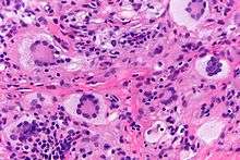
Touton giant cell
Touton giant cells are a type of multinucleated giant cell seen in lesions with high lipid content such as fat necrosis, xanthoma, and xanthelasma and xanthogranulomas.They are also found in dermatofibroma.[1]

History
Touton giant cells are named for Karl Touton, a German botanist and dermatologist.[2] Karl Touton first observed these cells in 1885 and named them "xanthelasmatic giant cells", a name which has since fallen out of favor.[3]
Appearance
Touton giant cells, being multinucleated giant cells, can be distinguished by the presence of several nuclei in a distinct pattern. They contain a ring of nuclei surrounding a central homogeneous cytoplasm, while foamy cytoplasm surrounds the nuclei.[4][5] The cytoplasm surrounded by the nuclei has been described as both amphophilic and eosinophilic, while the cytoplasm near the periphery of the cell is pale and foamy in appearance.[6]
Causes
Touton giant cells are formed by the fusion of macrophage-derived foam cells. It has been suggested that cytokines such as interferon gamma, interleukin-3, and M-CSF may be involved in the production of Touton giant cells.[7][8]
References
- Rapini, Ronald P.; Bolognia, Jean L.; Jorizzo, Joseph L. (2007). Dermatology: 2-Volume Set. St. Louis: Mosby. pp. 14, 15. ISBN 978-1-4160-2999-1.
- Aterman, K.; Remmele, W.; Smith, M. (Jun 1988). "Karl Touton and his "xanthelasmatic giant cell." A selective review of multinucleated giant cells". The American Journal of Dermatopathology. 10 (3): 257–269. ISSN 0193-1091. PMID 3068999.
- Olson, James (1989). The History of Cancer: An Annotated Bibliography. ABC-CLIO. p. 139. ISBN 9780313258893.
- Grant-Kels, Jane (2007). Color Atlas of Dermatopathology. City: Informa Healthcare. pp. 107, 119. ISBN 978-0-8493-3794-9.
- Carmen Gómez-Mateo, Maria; Monteagudo, Carlos (2013). "Nonepithelial skin tumors with multinucleated giant cells". Seminars in Diagnostic Pathology. 30 (1): 58–72. doi:10.1053/j.semdp.2012.01.004. PMID 23327730.
- Sequeira, Fiona; Gandhi, Suneil (2012). "Named cells in dermatology". Indian Journal of Dermatology, Venereology and Leprology. 78 (2): 207–16. doi:10.4103/0378-6323.93650. PMID 22421663.
- Quinn, Mark; Schepetkin, Igor (2009). "Role of NADPH Oxidase in Formation and Function of Multinucleated Giant Cells". Journal of Innate Immunology. 1 (6): 509–26. doi:10.1159/000228158. PMC 2919507. PMID 20375608.
- Reiss, AB; Patel, CA (2004). "Interferon-gamma impedes reverse cholesterol transport and promotes foam cell transformation in THP-1 human monocytes/macrophages". Medical Science Monitor. 10 (11): 420–5. PMID 15507847.