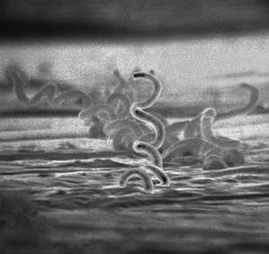
Spiral bacteria
Spiral bacteria, bacteria of spiral (helical) shape, form the third major morphological category of prokaryotes along with the rod-shaped bacilli and round cocci.[1][2] Spiral bacteria can be subclassified by the number of twists per cell, cell thickness, cell flexibility, and motility. The two types of spiral cells are spirillum and spirochete, with spirillum being rigid with external flagella, and spirochetes being flexible with internal flagella.[3]
Spirillum

A spirillum (plural spirilla) is a rigid spiral bacterium that is Gram-negative and frequently has external amphitrichous or lophotrichous flagella.[3] Examples include:
- Members of the genus Spirillum
- Campylobacter species, such as Campylobacter jejuni, a foodborne pathogen that causes campylobacteriosis
- Helicobacter species, such as Helicobacter pylori, a cause of peptic ulcers
Spirochete

A spirochete (plural spirochetes) is a very thin, elongate, flexible, spiral bacteria that is motile via internal periplasmic flagella inside the outer membrane.[3] They comprise the phylum Spirochaetes. Owing to their morphological properties, spirochetes are difficult to Gram-stain but may be visualized using dark field microscopy or Warthin–Starry stain.[4] Examples include:
- Leptospira species, which cause leptospirosis.
- Borrelia species, such as Borrelia burgdorferi, a tick-borne bacterium that causes Lyme disease
- Treponema species, such as Treponema pallidum, subspecies of which causes treponematoses, including syphilis
References
- Csuros, Maria; Csuros, Csaba (1999). Microbiological Examination of Water and Wastewater. Boca Raton, Florida: CRC Press. pp. 16–17. ISBN 9781566701792.
- Young, Kevin D. (September 2006). "The Selective Value of Bacterial Shape". Microbiology and Molecular Biology Reviews. 70 (3): 660–703. doi:10.1128/MMBR.00001-06. PMC 1594593. PMID 16959965.
- Taro, Kathleen (2007). Foundations in Microbiology (6th International ed.). McGraw-Hill. pp. 108–109. ISBN 0071262326. Retrieved 11 September 2017.
- Humphrey, Peter A.; Dehner, Louis P.; Pfeifer, John D., eds. (2008). "Chapter 53: Histology and histochemical stains". The Washington Manual of Surgical Pathology. Philadelphia: Lippincott Williams & Wilkins. p. 680. ISBN 9780781765275.