Nissl body
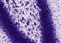
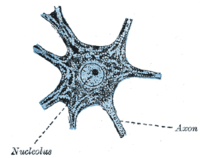
A Nissl body, also known as Nissl substance and Nissl material, is a large granular body found in neurons. These granules are of rough endoplasmic reticulum (RER) with rosettes of free ribosomes, and are the site of protein synthesis.[1] It was named after Franz Nissl, a German neuropathologist who invented the Nissl staining method.[2]


Nissl bodies can be demonstrated by a method of selective staining developed by Nissl (Nissl staining), using an aniline stain to label extranuclear RNA granules. This staining method is useful to localize the cell body, as it can be seen in the soma and dendrites of neurons, though not in the axon or axon hillock.[3] Due to RNA's basophilic ("base-loving") properties it is stained blue by this method.
Nissl bodies show changes under various physiological conditions and in pathological conditions they may dissolve and disappear (chromatolysis).
Function
The functions of Nissl bodies is thought to be the same as that of the rest of the ER and the Golgi apparatus: the manufacture and release of proteins and amino acids.[2]
The ultrastructure of Nissl bodies suggests they are primarily concerned with the synthesis of proteins for intracellular use.[4]
References
- John H. Byrne; James Lewis Roberts (23 January 2009). From Molecules to Networks: An Introduction to Cellular and Molecular Neuroscience. Academic Press. pp. 20–. ISBN 978-0-12-374132-5. Retrieved 4 January 2013.
- Richard H. Thompson (29 March 2000). The Brain: A Neuroscience Primer. Macmillan. pp. 35–. ISBN 978-0-7167-3226-6. Retrieved 4 January 2013.
- Wolfgang Kühnel (2003). Color Atlas of Cytology, Histology, and Microscopic Anatomy. Thieme. pp. 182–. ISBN 978-3-13-562404-4. Retrieved 4 January 2013.
- T. Herdegen; J. Delgado-Garcia (25 May 2005). Brain Damage and Repair: From Molecular Research to Clinical Therapy. Springer. pp. 37–. ISBN 978-1-4020-1892-3. Retrieved 4 January 2013.
External links

- Nissl+Bodies at the US National Library of Medicine Medical Subject Headings (MeSH)
- Histology image: 04103loa – Histology Learning System at Boston University - "Nervous Tissue and Neuromuscular Junction: spinal cord, cell bodies of anterior horn cells"
- Histology at anhb.uwa.edu.au
- Tissues containing Nissl bodies at harvard.edu