Trapezoid bone
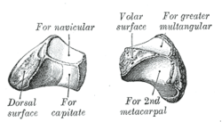
The trapezoid bone (lesser multangular bone) is a carpal bone in tetrapods, including humans. It is the smallest bone in the distal row of carpal bones that give structure to the palm of the hand. It may be known by its wedge-shaped form, the broad end of the wedge constituting the dorsal, the narrow end the palmar surface; and by its having four articular facets touching each other, and separated by sharp edges. It is homologous with the "second distal carpal" of reptiles and amphibians.
| Trapezoid bone | |
|---|---|
_01_palmar_view.png) Left hand anterior view (palmar view). Trapezoid bone shown in red. | |
 The left trapezoid bone. | |
| Details | |
| Articulations | articulates with four bones: scaphoid proximally second metacarpal distally trapezium bone laterally capitate medially |
| Identifiers | |
| Latin | os trapezoideum, os multangulum minus |
| MeSH | D051223 |
| TA | A02.4.08.010 |
| FMA | 23724 |
| Anatomical terms of bone | |
Structure
The trapezoid is a four-sided carpal bone found within the hand. The trapezoid is found within the distal row of carpal bones.[1] :708
Surfaces
The superior surface, quadrilateral, smooth, and slightly concave, articulates with the scaphoid.
The inferior surface articulates with the proximal end of the second metacarpal bone; it is convex from side to side, concave from before backward and subdivided by an elevated ridge into two unequal facets.
The dorsal and palmar surfaces are rough for the attachment of ligaments, the former being the larger of the two.
The lateral surface, convex and smooth, articulates with the trapezium.
The medial surface is concave and smooth in front, for articulation with the capitate; rough behind, for the attachment of an interosseous ligament.
Clinical Significance
Isolated fractures of the trapezoid are rare, representing 0.4% of the total, thus being the least common of all carpal fractures. This is due to the bone being in a fairly protected position. Distally, it forms a stable, relatively immobile joint with the second metacarpal, radially and proximally it forms strong ligaments with the trapezium and the capitate ulnarly, scaphoid respectively.
However, injury can occur through axial force applied to the second metacarpal base. Subluxations, such as ones caused by delivering a blow, are not uncommon. Direct trauma to the bone can also cause fracture.
Due to its rarity, standard treatment has not been established. A wide range of treatments are possible, including rest, surgery and casting.[2]
History
The etymology derives from the Greek trapezion which means "irregular quadrilateral," from tra- "four" and peza "foot" or "edge." Literally, "a little table" from trapeza meaning "table" and -oeides "shaped."
Additional images
_-_animation01.gif) Position of trapezoid bone (shown in red). Left hand. Animation.
Position of trapezoid bone (shown in red). Left hand. Animation._-_animation02.gif) Trapezoid bone of the left hand. Close up. Animation.
Trapezoid bone of the left hand. Close up. Animation. Trapezoid bone.
 Right hand posterior view (dorsal view). Thumb on bottom.
 Trapezoid shown in yellow. Left hand. Dorsal surface.
 Trapezoid shown in yellow. Left hand. Palmar surface.
 Transverse section across the wrist (palm on top, thumb on left). Trapezoid bone shown in yellow (labelled as "Lesser Multang").
 Cross section of wrist (thumb on left). Trapezoid shown in red (labelled as "Lesser Multang").
References
This article incorporates text in the public domain from page 225 of the 20th edition of Gray's Anatomy (1918)
- Drake, Richard L.; Vogl, Wayne; Tibbitts, Adam W.M. Mitchell; illustrations by Richard; Richardson, Paul (2005). Gray's anatomy for students. Philadelphia: Elsevier/Churchill Livingstone. ISBN 978-0-8089-2306-0.
- Sadowski, RM; Montilla, RD (2008). "Rare isolated trapezoid fracture: a case report". Hand (N Y). 3: 372–4. doi:10.1007/s11552-008-9100-8. PMC 2584218. PMID 18780025.