Radial tuberosity
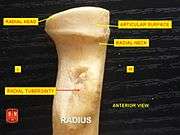
Beneath the neck of the radius, on the medial side, is an eminence, the radial tuberosity; its surface is divided into:
- a posterior, rough portion, for the insertion of the tendon of the biceps brachii.
- an anterior, smooth portion, on which a bursa is interposed between the tendon and the bone.
| Radial tuberosity | |
|---|---|
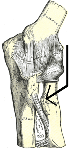
 Left elbow-joint, showing anterior and ulnar collateral ligaments. (Radial tuberosity visible at center right.) | |
 Bones of left forearm. Anterior aspect. (Radius is bone on right. Radial tuberosity is visible at upper left of radius.) | |
| Details | |
| Identifiers | |
| Latin | Tuberositas radii |
| TA | A02.4.05.007 |
| FMA | 23489 |
| Anatomical terms of bone | |
Sources
This article incorporates text in the public domain from page 219 of the 20th edition of Gray's Anatomy (1918)
gollark: Due to those, yes.
gollark: ++apioform
gollark: Unfortunately, this is not possible.
gollark: So I thought "what if I made the random stuff API secretly running 28% of osmarks.net even more convoluted by integrating a trivial LDAP server to just return some data from a config file, and also connecting this with the HTTP frontend for nginx subrequest authentication?".
gollark: However, OpenLDAP was very annoying.
External links
- Anatomy figure: 07:02-08 at Human Anatomy Online, SUNY Downstate Medical Center
- radiographsul at The Anatomy Lesson by Wesley Norman (Georgetown University) (xrayelbow)
Additional images
 Radial tuberosity shown.
 Anterior View. Radial tuberosity.
 Posterior View. Radial tuberosity.
 Radial tuberosity below neck.
 Radial tuberosity below neck.
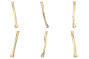
 Left Radius - close-up - animation.
.jpg) Elbow joint - deep dissection (anterior view, human cadaver)
Elbow joint - deep dissection (anterior view, human cadaver)- Elbow joint - deep dissection (anterior view, human cadaver)

This article is issued from Wikipedia. The text is licensed under Creative Commons - Attribution - Sharealike. Additional terms may apply for the media files.