Thecoma
Thecomas or theca cell tumors are benign ovarian neoplasms composed only of theca cells. Histogenetically they are classified as sex cord-stromal tumours.
| Thecoma | |
|---|---|
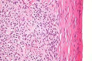 | |
| High magnification micrograph of a thecoma. H&E stain. | |
| Specialty | Oncology |
They are typically estrogen-producing and they occur in older women (mean age 59; 84% after menopause). (They can, however, appear before menopause.[1])
60% of patients present with abnormal uterine bleeding, and 20% have endometrial carcinoma.
Pathologic features
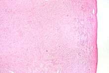
Low magnification micrograph of a thecoma showing compression of the ovarian cortex (right of image). H&E stain.
Grossly, the tumour is solid and yellow.
Grossly and microscopically, it consists of the ovarian cortex.
Microscopically, the tumour cells have abundant lipid-filled cytoplasm.
gollark: Although in our English lessons I generally ended up having time to reread the book a fwe times while we were doing it. So boring.
gollark: I never actually did do that. It probably would have saved time, in retrospect.
gollark: Unrelatedly, writing long things is hard and school has prepared me terribly for this.
gollark: Sounds fun *and* totally safe!
gollark: What's that log thing from?
References
- Okada I, Nakagawa S, Takemura Y, et al. (October 2004). "Ovarian thecoma associated in the first trimester of pregnancy". J. Obstet. Gynaecol. Res. 30 (5): 368–71. doi:10.1111/j.1447-0756.2004.00212.x. PMID 15327450.
This article is issued from Wikipedia. The text is licensed under Creative Commons - Attribution - Sharealike. Additional terms may apply for the media files.