Short posterior ciliary arteries
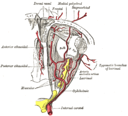
The short posterior ciliary arteries, around seven in number arise from the medial posterior ciliary artery and lateral posterior ciliary artery which are branches of the ophthalmic artery as it crosses the optic nerve.[1]
| Short posterior ciliary arteries | |
|---|---|
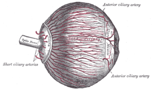
 The arteries of the choroid and iris. The greater part of the sclera has been removed. | |
 The ophthalmic artery and its branches. | |
| Details | |
| Source | Ophthalmic artery |
| Vein | Vorticose veins |
| Supplies | Choroid (up to the equator of the eye) ciliary processes |
| Identifiers | |
| Latin | Arteriae ciliares posteriores breves |
| TA | A12.2.06.031 |
| FMA | 70777 |
| Anatomical terminology | |
Course and target
They pass forward around the optic nerve to the posterior part of the eyeball, pierce the sclera around the entrance of the optic nerve, and supply the choroid (up to the equator of the eye) and ciliary processes.
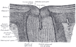
Some branches of the short posterior ciliary arteries also supply the optic disc via an anastomotic ring, the Circle of Zinn-Haller or Circle of Zinn, which is associated with the fibrous extension of the ocular tendons (Annulus of Zinn).
Additional images
 The terminal portion of the optic nerve and its entrance into the eyeball, in horizontal section.
gollark: It was quite long and most of it was unhelpful.
gollark: I see.
gollark: How DOES one utilize it?
gollark: Do I need to patch my kernel to fix this, then?
gollark: This *always* prints a status of 512. It should be not 512.
References
- Gray's anatomy : the anatomical basis of clinical practice. Standring, Susan (Forty-first ed.). [Philadelphia]. 2016. ISBN 978-0-7020-5230-9. OCLC 920806541.CS1 maint: others (link)
This article is issued from Wikipedia. The text is licensed under Creative Commons - Attribution - Sharealike. Additional terms may apply for the media files.