Lenticel
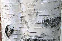
A lenticel is a porous tissue consisting of cells with large intercellular spaces in the periderm of the secondarily thickened organs and the bark of woody stems and roots of dicotyledonous flowering plants.[2] It functions as a pore, providing a pathway for the direct exchange of gases between the internal tissues and atmosphere through the bark, which is otherwise impermeable to gases. The name lenticel, pronounced with an [s], derives from its lenticular (lens-like) shape.[3] The shape of lenticels is one of the characteristics used for tree identification.[4]

Evolution
Before there was much evidence for the existence and functionality of lenticels, the fossil record has shown the first primary mechanism of aeration in early vascular plants to be the stomata.[5] However, in Woody plants, with vascular and cork cambial activity and secondary growth, the entire epidermis may be replaced by a suberized periderm or bark in which the functions of the stomata are replaced by lenticels.
The extinct arboreal plants of the genera Lepidodendron and Sigillaria were the first to have distinct aeration structures that rendered these modifications. "Parichnoi" (singular: parichnos) are canal-like structures that, in association with foliar traces of the stem, connected the stem's outer and middle cortex to the mesophyll of the leaf. Parichnoi were thought to eventually give rise to lenticels as they helped solve the issue of long-range oxygen transport in these woody plants during the Carboniferous period. They also acquired secondary connections as they evolved to become transversely elongated to efficiently aerate the maximum number of vertical rays as well as the central core tissue of the stem.[6] The evolutionary significance of parichnoi was their functionality in the absence of cauline stomata, where they can also be affected and destroyed by pressure similar to what can damage to stomatal tissue. Evidently, in both conifers and Lepidodendroids, the parichnoi, as the primary lenticular structure, appear as paired structures on either side of leaf scars. The development and increase in the number of these primitive lenticels were key to providing a system that was open for aeration and gas exchange in these plants.[7]
Structure and development
In plant bodies that produce secondary growth, lenticels promote gas exchange of oxygen, carbon dioxide, and water vapor.[8] Lenticel formation usually begins beneath stomatal complexes during primary growth preceding the development of the first periderm.The formation of lenticels seem to be directly related to the growth and strength of the shoot and on the hydrose of the tissue, which refers to the internal moisture.[9] As stems and roots mature lenticel development continues in the new periderm (for example, periderm that forms at the bottom of cracks in the bark).
Lenticels are found as raised circular, oval, or elongated areas on stems and roots. In woody plants, lenticels commonly appear as rough, cork-like structures on young branches. Underneath them, porous tissue creates a number of large intercellular spaces between cells. This tissue fills the lenticel and arises from cell division in the phellogen or substomatal ground tissue. Discoloration of lenticels may also occur, such as in mangoes, that may be due to the amount of lignin in cell walls.[10][11]
In oxygen deprived conditions, making respiration a daily challenge, different species may possess specialized structures where lenticels can be found. For example, in a common mangrove species, lenticels appear on pneumatophores (specialized roots), where the parenchyma cells that connect to the aerenchyma structure increase in size and go through cell division.[12] In contrast, lenticels in grapes are located on the pedicels and act as a function of temperature. If they are blocked, hypoxia and successive ethanol accumulation may result and lead to cell death.[13]
Fruits

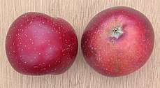
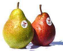
Lenticels are also present on many fruits, quite noticeably on many apples and pears. On European pears, they can serve as an indicator of when to pick the fruit, as light lenticels on the immature fruit darken and become brown and shallow from the formation of cork cells[14][15] Certain bacterial and fungal infections can penetrate fruits through their lenticels, with susceptibility sometimes increasing with its age.[16]
As mentioned previously, the term lenticel is usually associated with the breakage of periderm tissue that is associated with gas exchange; however, lenticels also refer to the lightly colored spots found on apples (a type of pome fruit). "Lenticel" seems to be the most appropriate term to describe both structures mentioned in light of their similar function in gas exchange. Pome lenticels can be derived from (1) no longer functioning stomata, (2) epidermal breaks from the removal of trichomes, and (3) other epidermal breaks that usually occur in the early development of young pome fruits. The closing of pome lenticels can arise when the cuticle over the stomata opening or the substomatal layer seals. Closing can also begin if the substomatal cells become suberized, like cork. The number of lenticels usually varies between the species of apples, where the range may be from 450 to 800 or from 1500 to 2500 in Winesap and Spitzenburg apples, respectively. This wide range may be due to the water availability during the early stages of development of each apple type.[17]
“Lenticel breakdown” is a global skin disorder of apples in which lenticels develop dark 1–8 mm diameter pits shortly after processing and packing.[18][19] It is most common on the ‘Gala’ (Malus × domestica) variety, particularly the ‘Royal Gala’, and also occurs in ‘Fuji’, ‘Granny Smith’, ‘Golden Delicious’, and ‘Delicious’ varieties.[18][19] It is more common in arid regions, and is thought to be related to relative humidity and temperature.[18][19] The effect can be mitigated by spraying the fruit with lipophilic coatings prior to harvest.[18]
Tubers
Lenticels are also present on potato tubers.[20]
Gallery
- Lenticels on Prunus serrula
- Lenticels on Wild Cherry or Gean.
- Alder bark (Alnus glutinosa) with characteristic lenticels and abnormal lenticels on callused areas.
 Lenticels on potatoes of the Monalisa variety.
 Lenticels on Williams pear varieties.
Notes
- "Lenticel". The American Heritage Science Dictionary, Houghton Mifflin Company, via dictionary.com. Retrieved on 2007-10-11
- Gibson, Arthur C. "Bark Features in General Botany". Archived from the original on 2013-11-17. Retrieved 2012-08-19.
- Esau, K. (1953), Plant Anatomy, John Wiley & Sons Inc. New York, Chapman & Hall Ltd. London
- Michael G. Andreu; Erin M. Givens; Melissa H. Friedman what the. "How to Identify a Tree". University of Florida IFAS extension. Archived from the original on 2013-05-14. Retrieved 2013-03-07.
- Matthews, Jack S.A.; Vialet-Charbrand, Silvere R.M.; Lawson, Tracy (June 2017). "Open Access Diurnal Variation in Gas Exchange: The Balance between Carbon Fixation and Water Loss". Plant Physiology. 174 (2): 614–623. doi:10.1104/pp.17.00152. PMC 5462061. PMID 28416704.
- Wetmore, Ralph H. (September 1926). "Organization and Significance of Lenticels in Dicotyledons. I. Lenticels in Relation to Aggregate and Compound Storage Rays in Woody Stems. Lenticels and Roots". Botanical Gazette. 82 (1): 71–78. doi:10.1086/333634. JSTOR 2470894.
- Hook, Donald D. (December 1972). "Aeration in Trees". Botanical Gazette. 133 (4): 443–454. doi:10.1086/336669. JSTOR 2474119.
- Lendzian, Klaus J. (July 4, 2006). "Survival strategies of plants during secondary growth: barrier properties of phellems and lenticels towards water, oxygen, and carbon dioxide". Experimental Botany. 57 (11): 2535–2546. doi:10.1093/jxb/erl014. PMID 16820395.
- Priestley, J. H. (December 18, 1922). "Physiological Studies in Plant Anatomy V. Causal Factors in Cork Formation". The New Phytologist. 21 (5): 252–268. doi:10.1111/j.1469-8137.1922.tb07603.x. JSTOR 2428084.
- Kenoyer, Leslie A. (Dec. 31, 1908 – Jan. 2, 1909). "Winter Condition of Lenticels". Transactions of the Kansas Academy of Science. 22: 323–326. doi:10.2307/3624744. JSTOR 3624744.CS1 maint: date format (link)
- Tamjinda, Boonchai (1992). "Anatomy of Lenticels and The Occurrence of Their Discoloration in Mangoes" (PDF). Natural Sciences. 26 (Suppl 5): 57–64.
- Purnobasuki, Hery (January 2011). "Structure of Lenticels on the Pneumatophores of Avicennia marina: as Aerating Device Deliver Oxygen in Mangrove's root". Biota. 16 (2): 309–315. doi:10.24002/biota.v16i2.113.
- Xiao, Zeyu; Rogiers, Suzy Y.; Sadras, Victor O.; Tyerman, Stephen D. (2018). "Hypoxia in grape berries: the role of seed respiration and lenticels on the berry pedicel and the possible link to cell death". Experimental Botany. 69 (8): 2071–2083. doi:10.1093/jxb/ery039. PMC 6018838. PMID 29415235.
- Krewer, Gerard; Bertrand, Paul. "Home garden pears". CAES Publications. University of Georgia College of Agricultural and Environmental Sciences. Retrieved 18 January 2014.
- Pyzner, John (19 April 2005). "Pick pears before completely ripe, advises LSU AgCenter horticulturist". Louisiana State University Agricultural Center website. Archived from the original on 15 January 2006.
- Irtwange, S. V. (February, 2006.) "Application of modified atmosphere packaging and related technology in postharvest handling of fresh fruits and vegetables" Archived 2006-10-11 at the Wayback Machine. Agricultural Engineering International: the CIGR Ejournal. Invited Overview No. 4. Vol. VIII, page 8. Retrieved on 2007-10-11.
- Clements, Harry F. (September 1935). "Morphology and Physiology of the Pome Lenticels of Pyrus malus". Botanical Gazette. 97 (1): 101–117. doi:10.1086/334539. JSTOR 2471695.
- Curry, Eric A.; Torres, Carolina; Neubauer, Luis (2008). "Preharvest lipophilic coatings reduce lenticel breakdown disorder in 'Gala' apples". HortTechnology. 18 (4): 690–696. doi:10.21273/HORTTECH.18.4.690.
- Turketti, S.S.; Curry, E.; Lötze, E. (2012). "Role of lenticel morphology, frequency and density on incidence of lenticel breakdown in 'Gala' apples". Scientia Horticulturae. 138: 90–95. doi:10.1016/j.scienta.2012.02.010. ISSN 0304-4238.
- Adams, M. J. (1975). "Potato tuber lenticels: Development and structure". Annals of Applied Biology. 79 (3): 265–273. doi:10.1111/j.1744-7348.1975.tb01582.x.
References
- Raven, Peter H.; Ray F. Evert; Susan E. Eichorn (2005). Biology of Plants 7th Ed. W.H. Freeman and Company Publishers. pp. 586–587. ISBN 0-7167-1007-2.