Sacral plexus
In human anatomy, the sacral plexus is a nerve plexus which provides motor and sensory nerves for the posterior thigh, most of the lower leg and foot, and part of the pelvis. It is part of the lumbosacral plexus and emerges from the lumbar vertebrae and sacral vertebrae (L4-S4).[1] A sacral plexopathy is a disorder affecting the nerves of the sacral plexus, usually caused by trauma, nerve compression, vascular disease, or infection. Symptoms may include pain, loss of motor control, and sensory deficits.
| Sacral plexus | |
|---|---|
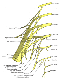 Plan of sacral and pudendal plexuses. | |
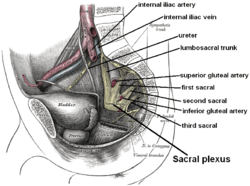 Relations of the sacral plexus. Dissection of side wall of pelvis showing sacral and pudendal plexuses. | |
| Details | |
| From | L4-L5, S1-S4 |
| Identifiers | |
| Latin | plexus sacralis |
| TA | A14.2.07.027 |
| FMA | 5909 |
| Anatomical terms of neuroanatomy | |
Structure
The sacral plexus is formed by:
- the lumbosacral trunk
- the anterior division of the first sacral nerve
- portions of the anterior divisions of the second and third sacral nerves
The nerves forming the sacral plexus converge toward the lower part of the greater sciatic foramen, and unite to form a flattened band, from the anterior and posterior surfaces of which several branches arise. The band itself is continued as the sciatic nerve, which splits on the back of the thigh into the tibial nerve and common fibular nerve; these two nerves sometimes arise separately from the plexus, and in all cases their independence can be shown by dissection. Often, the sacral plexus and the lumbar plexus are considered to be one large nerve plexus, the lumbosacral plexus. The lumbosacral trunk connects the two plexuses.
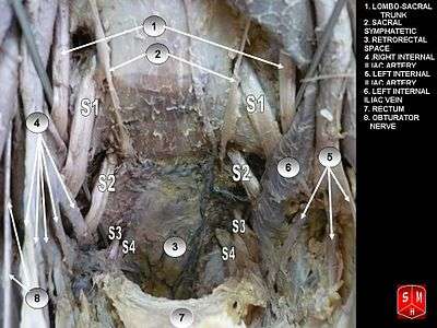
Relations
The sacral plexus lies on the back of the pelvis in front of the piriformis muscle and the pelvic fascia. In front of it are the internal iliac artery, internal iliac vein, the ureter, and the sigmoid colon. The superior gluteal artery and vein run between the lumbosacral trunk and the first sacral nerve, and the inferior gluteal artery and vein between the second and third sacral nerves.
Nerves formed
All the nerves entering the plexus, with the exception of the third sacral, split into ventral and dorsal divisions, and the nerves arising from these are as follows of the table below:
| Nerve | Segment | Innervated muscles | Cutaneous branches |
|---|---|---|---|
| Superior gluteal | L4-S1 | Gluteus medius Gluteus minimus Tensor fasciae latae |
|
| Inferior gluteal | L5-S2 | Gluteus maximus | |
| Posterior cutaneous femoral | S1-S3 |
Posterior cutaneous femoral | |
| Direct branches from plexus | |||
|
S1-2 | Piriformis | |
|
L5, S1-2 | Obturator internus and Superior gemellus | |
| L4-5, S1 | Quadratus femoris and Inferior gemellus | ||
| Sciatic | |||
| Sciatic | L4-S3 | Semitendinosus (Tib) Semimembranosus (Tib) Biceps femoris
Adductor magnus (medial part, Tib) |
|
| Common fibular | L4-S2 | Lateral sural cutaneous Communicating fibular | |
|
Peroneus longus Peroneus brevis |
Medial dorsal cutaneous Intermediate dorsal cutaneous | |
|
Tibialis anterior Extensor digitorum longus Extensor digitorum brevis Extensor hallucis longus Extensor hallucis brevis Peroneus tertius |
Lateral cutaneous nerve of big toe Intermediate dorsal cutaneous | |
| Tibial nerve | L4-S3 | Triceps surae Plantaris Popliteus Tibialis posterior Flexor digitorum longus Flexor hallucis longus |
Medial sural cutaneous Lateral calcaneal Medial calcaneal Lateral dorsal cutaneous |
| Abductor hallucis Flexor digitorum brevis Flexor hallucis brevis (medial head) Lumbrical (first and second) |
Proper digital plantar | ||
| Flexor hallucis brevis (lateral head) Quadratus plantae Abductor digiti minimi Flexor digiti minimi Lumbrical (third and fourth) Plantar interossei (first to third) Dorsal interossei (first to fifth) Adductor hallucis |
Proper plantar digital | ||
| Pudendal and coccygeal | |||
| Pudendal (Pudendal plexus) |
S2-S4 | Muscles of the pelvic floor: Levator ani Superficial transverse perineal Deep transverse perineal Bulbospongiosus Ischiocavernosus Sphincter anus externus Urethral sphincter |
Inferior rectal Perineal
|
| Coccygeal (Coccygeal plexus) |
S5-Co1 | Coccygeus | Anococcygeal Dorsal branches |
Additional images
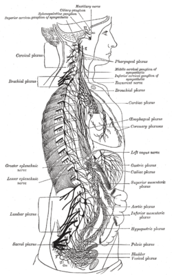 The right sympathetic chain and its connections with the thoracic, abdominal, and pelvic plexuses.
 A schematic depiction.
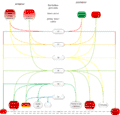 diagram of the Sacral plexus
See also
Notes
- Thieme Atlas of Anatomy (2006), pp 470-471
- Thieme Atlas of Anatomy (2006), pp 476, 478, 482
References
This article incorporates text in the public domain from page 957 of the 20th edition of Gray's Anatomy (1918)
- Thieme Atlas of Anatomy: General Anatomy and Musculoskeletal System. Thieme. 2006. ISBN 1-58890-419-9.
External links
| Wikimedia Commons has media related to Sacral plexus. |
- Lumbosacral+Plexus at the US National Library of Medicine Medical Subject Headings (MeSH)
- Cross section image: pembody/body15a—Plastination Laboratory at the Medical University of Vienna
- MedicalMnemonics.com: 3544 2382
- Illustration at backpain-guide.com
| Authority control |
|---|