Stable isotope labeling by amino acids in cell culture
Stable Isotope Labeling by/with Amino acids in Cell culture (SILAC) is a technique based on mass spectrometry that detects differences in protein abundance among samples using non-radioactive isotopic labeling.[1][2][3][4] It is a popular method for quantitative proteomics.

Procedure
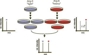
Two populations of cells are cultivated in cell culture. One of the cell populations is fed with growth medium containing normal amino acids. In contrast, the second population is fed with growth medium containing amino acids labeled with stable (non-radioactive) heavy isotopes. For example, the medium can contain arginine labeled with six carbon-13 atoms (13C) instead of the normal carbon-12 (12C). When the cells are growing in this medium, they incorporate the heavy arginine into all of their proteins. Thereafter, all peptides containing a single arginine are 6 Da heavier than their normal counterparts. Alternatively, uniform labeling with 13C or 15N can be used. The trick is that the proteins from both cell populations can be combined and analyzed together by mass spectrometry. Pairs of chemically identical peptides of different stable-isotope composition can be differentiated in a mass spectrometer owing to their mass difference. The ratio of peak intensities in the mass spectrum for such peptide pairs reflects the abundance ratio for the two proteins.[5][3]
Applications
A SILAC approach involving incorporation of tyrosine labeled with nine carbon-13 atoms (13C) instead of the normal carbon-12 (12C) has been utilized to study tyrosine kinase substrates in signaling pathways.[6] SILAC has emerged as a very powerful method to study cell signaling, post translation modifications such as phosphorylation,[6][7] protein–protein interaction and regulation of gene expression. In addition, SILAC has become an important method in secretomics, the global study of secreted proteins and secretory pathways.[8] It can be used to distinguish between proteins secreted by cells in culture and serum contaminants.[9] Standardized protocols of SILAC for various applications have also been published.[10][11]
While SILAC had been mostly used in studying eukaryotic cells and cell cultures, it had been recently employed in bacteria and its multicellular biofilm in antibiotic tolerance, to differentiate tolerance and sensitive subpopulations.[12]
Pulsed SILAC
Pulsed SILAC (pSILAC) is a variation of the SILAC method where the labelled amino acids are added to the growth medium for only a short period of time. This allows monitoring differences in de novo protein production rather than raw concentration.[13]
It had also been used to study biofilm tolerance to antibiotics to differentiate tolerant and sensitive subpopulations [12]
NeuCode SILAC
Traditionally the level of multiplexing in SILAC was limited due to the number of SILAC isotopes available. Recently, a new technique called NeuCode (neutron encoding) SILAC, has augmented the level of multiplexing achievable with metabolic labeling (up to 4).[14] The NeuCode amino acid method is similar to SILAC but differs in that the labeling only utilizes heavy amino acids. The use of only heavy amino acids eliminates the need for 100% incorporation of amino acids needed for SILAC. The increased multiplexing capability of NeuCode amino acids is from the use of mass defects from extra neutrons in the stable isotopes. These small mass differences however need to be resolved on high resolution mass spectrometers.
References
- Oda Y, Huang K, Cross FR, Cowburn D, Chait BT (June 1999). "Accurate quantitation of protein expression and site-specific phosphorylation". Proc. Natl. Acad. Sci. U.S.A. 96 (12): 6591–6. Bibcode:1999PNAS...96.6591O. doi:10.1073/pnas.96.12.6591. PMC 21959. PMID 10359756.
- Jiang H, English AM (2002). "Quantitative analysis of the yeast proteome by incorporation of isotopically labeled leucine". J. Proteome Res. 1 (4): 345–50. doi:10.1021/pr025523f. PMID 12645890.
- Ong SE, Blagoev B, Kratchmarova I, Kristensen DB, Steen H, Pandey A, Mann M (May 2002). "Stable isotope labeling by amino acids in cell culture, SILAC, as a simple and accurate approach to expression proteomics". Mol. Cell. Proteomics. 1 (5): 376–86. doi:10.1074/mcp.M200025-MCP200. PMID 12118079.
- Zhu H, Pan S, Gu S, Bradbury EM, Chen X (2002). "Amino acid residue specific stable isotope labeling for quantitative proteomics". Rapid Commun. Mass Spectrom. 16 (22): 2115–23. Bibcode:2002RCMS...16.2115Z. doi:10.1002/rcm.831. PMID 12415544.
- Schoeters, Floris; Van Dijck, Patrick (2019). "Protein-Protein Interactions in Candida albicans". Frontiers in Microbiology. 10: 1792. doi:10.3389/fmicb.2019.01792. ISSN 1664-302X. PMC 6693483. PMID 31440220.
- Ibarrola N, Molina H, Iwahori A, Pandey A (April 2004). "A novel proteomic approach for specific identification of tyrosine kinase substrates using [13C]tyrosine". J. Biol. Chem. 279 (16): 15805–13. doi:10.1074/jbc.M311714200. PMID 14739304.
- Ibarrola N, Kalume DE, Gronborg M, Iwahori A, Pandey A (November 2003). "A proteomic approach for quantitation of phosphorylation using stable isotope labeling in cell culture". Anal. Chem. 75 (22): 6043–9. doi:10.1021/ac034931f. PMID 14615979.
- Hathout Y (April 2007). "Approaches to the study of the cell secretome". Expert Rev Proteomics. 4 (2): 239–48. doi:10.1586/14789450.4.2.239. PMID 17425459.
- Polacek, Martin; Bruun, Jack-Ansgar; Johansen, Oddmund; Martinez, Inigo (2010). "Differences in the secretome of cartilage explants and cultured chondrocytes unveiled by SILAC technology". Journal of Orthopaedic Research. 28 (8): 1040–9. doi:10.1002/jor.21067. PMID 20108312.
- Amanchy R, Kalume DE, Pandey A (January 2005). "Stable isotope labeling with amino acids in cell culture (SILAC) for studying dynamics of protein abundance and posttranslational modifications". Sci. STKE. 2005 (267): pl2. doi:10.1126/stke.2672005pl2. PMID 15657263.
- Harsha HC, Molina H, Pandey A (2008). "Quantitative proteomics using stable isotope labeling with amino acids in cell culture". Nat Protoc. 3 (3): 505–16. doi:10.1038/nprot.2008.2. PMID 18323819.
- Chua SL, Yam JK, Sze KS, Yang L (2016). "Selective labelling and eradication of antibiotic-tolerant bacterial populations in Pseudomonas aeruginosa biofilms". Nat Commun. 7: 10750. Bibcode:2016NatCo...710750C. doi:10.1038/ncomms10750. PMC 4762895. PMID 26892159.
- Schwanhäusser B, Gossen M, Dittmar G, Selbach M (January 2009). "Global analysis of cellular protein translation by pulsed SILAC". Proteomics. 9 (1): 205–9. doi:10.1002/pmic.200800275. PMID 19053139.
- Merrill AE, Hebert AS, MacGilvray ME, Rose CM, Bailey DJ, Bradley JC, Wood WW, El Masri M, Westphall MS, Gasch AP, Coon JJ (September 2014). "NeuCode labels for relative protein quantification". Mol. Cell. Proteomics. 13 (9): 2503–12. doi:10.1074/mcp.M114.040287. PMC 4159665. PMID 24938287.
Further reading
- Ong SE, Kratchmarova I, Mann M (2003). "Properties of 13C-substituted arginine in stable isotope labeling by amino acids in cell culture (SILAC)". Journal of Proteome Research. 2 (2): 173–81. doi:10.1021/pr0255708. PMID 12716131.
- Ong SE, Mann M (2006). "A practical recipe for stable isotope labeling by amino acids in cell culture (SILAC)". Nature Protocols. 1 (6): 2650–60. doi:10.1038/nprot.2006.427. PMID 17406521.
- Ong SE, Mann M (2007). Stable isotope labeling by amino acids in cell culture for quantitative proteomics. Methods in Molecular Biology. 359. pp. 37–52. doi:10.1007/978-1-59745-255-7_3. ISBN 978-1-58829-571-2. PMID 17484109.