SHC1
SHC-transforming protein 1 is a protein that in humans is encoded by the SHC1 gene.[5] SHC has been found to be important in the regulation of apoptosis and drug resistance in mammalian cells.
SCOP classifies the 3D structure as belonging to the SH2 domain family.
Gene and expression
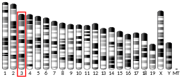
The gene SHC1 is located on chromosome 1 and encodes 3 main protein isoforms: p66SHC, p52SHC and p46SHC. These proteins differ in activity and subcellular locations, p66 is the longest and while the p52 and p46 link activated receptor tyrosine kinase to the RAS pathway.[6] The protein SHC1 also acts as a scaffold protein which is used in cell surface receptors.[7] The three proteins that SHC1 codes for have distinctly different molecular weights.[8] All three SHC1 proteins share the same domain arrangement consisting of an N-terminal phosphotyrosine-binding(PTB) domain and a C-terminal Src-homology2(SH2) domain. Both of the domains for the three proteins can bind to tyrosine-phosphorylated proteins but they are different in their phosphopeptide-binding specificities.[9] P66SHC is characterized by having an additional N-terminal CH2 domain.[9]
Function
Overexpression of SHC proteins are associated with cancer mitogenesis, carcinogenesis and metastasis.[8] The SHC and its adaptor proteins transmit signaling of the cell surface receptors such as EGFR, erbV-2 and insulin receptors. p52SHC and p46SHC activate the Ras-ERK pathway. p66SHC inhibits ERK1/2 activity and antagonize mitogenic and survival abilities of T-lymphoma Jurkat cell lines.[8] A rise in p66SHC promotes stress induced apoptosis.[8] p66SHC functionally is also involved in regulating oxidative and stress- induced apoptosis – mediating steroid action through the redox signaling pathway. P52SHC and p66SHC have been found in steroid hormone-regulated cancer and metastasizes.[8]
EGFR pathway
SHC1 has been found to act in signaling information after epidermal growth factor (EGF) stimulation. Activated tyrosine kinase receptors, on the cell surface, use proteins such as SHC1 that contain phosphotyrosine binding domains. After the EGF stimulation SHC1 binds to groups of proteins that activate survival pathways. This activation is followed by a sub-network of proteins that bind to SHC1 and are involved cytoskeleton reorganization, trafficking and signal termination. PTPN122 then acts as a switch to convert SHC1 to SgK269-mediated pathways that regulate cell invasion and morphogenesis.[7] SHC1 is not a static scaffold protein, a protein that does not move or change over time, it is dynamic as the conformation changes and modifies the EGFR signaling output over time.[10]
MCT-1 regulation
SHC proteins are differentially regulated by the Multiple Copies in T-cell malignancy(MCT-1). This regulation affects the SHC-Ras-ERK pathway.[8] With MCT-1 reduction the phosphor activation of Ras, MEK and ERk ½ were also reduced, this reduction in ERK also affects cyclin D1. The expression of the SHC proteins (all three) were also dramatically reduced with the reduction of MCT-1 because of this it is thought that MCT-1 acts as an inducer of SHC gene transcription. p66SHC is found to be the protein that is most affected by MCT-1. SHC expression downregulated in tumorigenic processes are identified after MCT-1 depletion. By blocking the MCT-1 activity this could inhibit the SHC signaling cascase and the oncogenicty and tumorigenicity that is regulated by SHC expression.[8]
Oxidative stress
Oxidative stress occurs when the production of reactive oxygen species (ROS) is greater than their catabolism. ROS production by the mitochondria is regulated by many diverse factors including SHC1.[11] The SHC proteins are regulated by tyrosine phosphorylation and are part of the growth factor and stress-induced ERK activation. There have been findings that suggest a correlation between life span and the oxidative stress response. Selective resistance to oxidative stress and extended life span have been related to p66SHC.[12]
Life span
There is a link between oxidative stress, life span and p66SHC[12] in mice because of this relationship the SHC gene has been related to longevity and increasing the life span of the mouse.[13] It has been proposed that SHC1 modulates the life span and stress response through the DAF-2 insulin- like receptor of the IIS pathway. The SHC-1 can directly interact with the DAF-2 in vitro.[9]
p66SHC metabolism
p66SHC operates as a redox enzyme linked to apoptotic cell death. p66SHC has been related to the sirtuin-1 system and has been associated with endothelial damage and repair. This relationships is also related to vascular homeostasis and oxidative stress.[14] p66SHC can be altered by changes in the glucose metabolism and vascular senescence. When protein kinase C is induced by hyperglycemia, p66SCH is induced which then leads to oxidative stress. When the coagulated protease-activated protein C inhibits p66SHC a cytoprotective effect on diabetic nephropathy is placed on the kidneys . When a mutations such as a p66SHC deletion occurs the cardiomyocyte death is reduced and a pool of cardiac stem cells are preserved from oxidative damage – preventing diabetic cardiomyopathy. The deletion of p66SHC also protects from ischemia/reperfusion brain injuries through blunted production of free radicals.[14]
Clinical significance
The signaling activation of SHC is implicated in tumorigenic in cancer cells there is a potential to use SHC as a prognostic marker when targeting cancer treatment.[8] SHC1 interacts with SgK269 which is a member of the Src kinase signaling network that characterized basal breast cancer cells. When SgK269 is overexpressed in mammary epithelial cells it promotes the cell growth and might contribute to the progression of aggressive breast cancers.[15] In prostate and ovarian cancer, increased expression of p66Shc appears to promote cell proliferation.[16] and tumorigenicity, particularly in prostate cancer xenografts[17] This tumorigenic effect is related to its ability to increase redox stress in these cancer cells.[18]
References
- GRCh38: Ensembl release 89: ENSG00000160691 - Ensembl, May 2017
- GRCm38: Ensembl release 89: ENSMUSG00000042626 - Ensembl, May 2017
- "Human PubMed Reference:". National Center for Biotechnology Information, U.S. National Library of Medicine.
- "Mouse PubMed Reference:". National Center for Biotechnology Information, U.S. National Library of Medicine.
- Pelicci G, Lanfrancone L, Grignani F, McGlade J, Cavallo F, Forni G, Nicoletti I, Grignani F, Pawson T, Pelicci PG (Jul 1992). "A novel transforming protein (SHC) with an SH2 domain is implicated in mitogenic signal transduction". Cell. 70 (1): 93–104. doi:10.1016/0092-8674(92)90536-L. PMID 1623525.
- "Genes and Mapped Phenotypes". National Center for Biotechnology Information. US National Library of Medicine.
- Zheng Y, Zhang C, Croucher DR, Soliman MA, St-Denis N, Pasculescu A, Taylor L, Tate SA, Hardy WR, Colwill K, Dai AY, Bagshaw R, Dennis JW, Gingras AC, Daly RJ, Pawson T (Jul 2013). "Temporal regulation of EGF signalling networks by the scaffold protein Shc1". Nature. 499 (7457): 166–71. doi:10.1038/nature12308. PMC 4931914. PMID 23846654.
- Shih HJ, Chen HH, Chen YA, Wu MH, Liou GG, Chang WW, Chen L, Wang LH, Hsu HL (Nov 2012). "Targeting MCT-1 oncogene inhibits Shc pathway and xenograft tumorigenicity". Oncotarget. 3 (11): 1401–15. doi:10.18632/oncotarget.688. PMC 3717801. PMID 23211466.
- Neumann-Haefelin E, Qi W, Finkbeiner E, Walz G, Baumeister R, Hertweck M (Oct 2008). "SHC-1/p52Shc targets the insulin/IGF-1 and JNK signaling pathways to modulate life span and stress response in C. elegans". Genes & Development. 22 (19): 2721–35. doi:10.1101/gad.478408. PMC 2559911. PMID 18832074.
- Wrighton KH (Aug 2013). "Cell signalling: EGF signalling--it's all in SHC1's timing". Nature Reviews Molecular Cell Biology. 14 (8): 463. doi:10.1038/nrm3630. PMID 23860237.
- Nathan C, Cunningham-Bussel A (May 2013). "Beyond oxidative stress: an immunologist's guide to reactive oxygen species". Nature Reviews. Immunology. 13 (5): 349–61. doi:10.1038/nri3423. PMC 4250048. PMID 23618831.
- Finkel T, Holbrook NJ (Nov 2000). "Oxidants, oxidative stress and the biology of ageing". Nature. 408 (6809): 239–47. doi:10.1038/35041687. PMID 11089981.
- Mooijaart SP, van Heemst D, Schreuder J, van Gerwen S, Beekman M, Brandt BW, Eline Slagboom P, Westendorp RG (Feb 2004). "Variation in the SHC1 gene and longevity in humans". Experimental Gerontology. 39 (2): 263–8. doi:10.1016/j.exger.2003.10.001. PMID 15036421.
- Avogaro A, de Kreutzenberg SV, Federici M, Fadini GP (Jun 2013). "The endothelium abridges insulin resistance to premature aging". Journal of the American Heart Association. 2 (3): e000262. doi:10.1161/JAHA.113.000262. PMC 3698793. PMID 23917532.
- Dikic I, Daly RJ (Mar 2012). "Signalling through the grapevine". EMBO Reports. 13 (3): 178–80. doi:10.1038/embor.2012.16. PMC 3323131. PMID 22354089.
- Bhat SS, Anand D, Khanday FA (2015). "p66Shc as a switch in bringing about contrasting responses in cell growth: implications on cell proliferation and apoptosis". Molecular Cancer. 14: 76. doi:10.1186/s12943-015-0354-9. PMC 4421994. PMID 25890053.
- Veeramani S, Chou YW, Lin FC, Muniyan S, Lin FF, Kumar S, Xie Y, Lele SM, Tu Y, Lin MF (July 2012). "Reactive oxygen species induced by p66Shc longevity protein mediate nongenomic androgen action via tyrosine phosphorylation signaling to enhance tumorigenicity of prostate cancer cells". Free Radical Biology & Medicine. 53 (1): 95–108. doi:10.1016/j.freeradbiomed.2012.03.024. PMC 3384717. PMID 22561705.
- Lebiedzinska-Arciszewska M, Oparka M, Vega-Naredo I, Karkucinska-Wieckowska A, Pinton P, Duszynski J, Wieckowski MR (2015). "The interplay between p66Shc, reactive oxygen species and cancer cell metabolism". European Journal of Clinical Investigation. 45 Suppl 1: 25–31. doi:10.1111/eci.12364. PMID 25524583.
Further reading
- Sasaoka T, Kobayashi M (Aug 2000). "The functional significance of Shc in insulin signaling as a substrate of the insulin receptor". Endocrine Journal. 47 (4): 373–81. doi:10.1507/endocrj.47.373. PMID 11075717.
- Ravichandran KS (Oct 2001). "Signaling via Shc family adapter proteins". Oncogene. 20 (44): 6322–30. doi:10.1038/sj.onc.1204776. PMID 11607835.
- van der Geer P (May 2002). "Phosphorylation of LRP1: regulation of transport and signal transduction". Trends in Cardiovascular Medicine. 12 (4): 160–5. doi:10.1016/S1050-1738(02)00154-8. PMID 12069755.


