Letterer–Siwe disease
Letterer–Siwe disease is one of the four recognized clinical syndromes of Langerhans cell histiocytosis (LCH). It causes approximately 10% of LCH disease and is the most severe form.[1] Prevalence is estimated at 1:500,000 and the disease almost exclusively occurs in children less than three years old.[2] The name is derived from the names of Erich Letterer and Sture Siwe.
| Letterer–Siwe disease | |
|---|---|
| Other names | Acute and disseminated Langerhans cell histiocytosis |
 | |
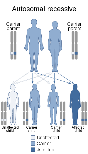
| This condition is inherited in an autosomal recessive manner | |
| Specialty | Oncology |
Presentation
Letterer-Siwe is characterized by skin lesions, ear drainage, lymphadenopathy, osteolytic lesions, and hepatosplenomegaly. The skin lesions are scaly and may involve the scalp, ear canals, and abdomen.[3]
Cause
Oncogenic mutation of BRAF 50-70% cases
Diagnosis
The diagnosis was established by histopathology and electron microscopy.--2. In Abt-Letterer-Siwe disease, the racket-like Langerhans cell granules are found by electron microscopy within the specific infiltrating cells. The demonstration of these organelles allows the unequivocal diagnosis in cases with uncharacteristic clinical or histopathological appearance. The same structures are characteristic of Hand-Schüller-Christian disease and of eosinophilic granuloma. The electron microscopic findings confirm the grouping of these three diseases together as "histiocytosis X".
Prognosis
The disease is often rapidly fatal, with a five-year survival rate of 50%. The development of thrombocytopenia is a poor prognostic sign.[1]
References
- "Langerhans Cell Histiocytosis - Hematology and Oncology - Merck Manuals Professional Edition". Merck Manuals Professional Edition. Retrieved 2017-05-19.
- RESERVED, INSERM US14 -- ALL RIGHTS. "Orphanet: Letterer Siwe disease". www.orpha.net. Retrieved 2017-05-19.
- "Langerhans cell histiocytosis | DermNet New Zealand". www.dermnetnz.org. Retrieved 2017-05-19.
External links
| Classification | |
|---|---|
| External resources |