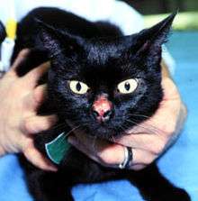
Eosinophilic granuloma
Eosinophilic granuloma is a form of Langerhans cell histiocytosis. It is a condition of both human and veterinary pathology.
| Eosinophilic granuloma | |
|---|---|
| Other names | Rodent ulcer |
 | |
| Eosinophilic granuloma | |
| Specialty | Dermatology |
Feline eosinophilic granuloma complex is synonymous with feline eosinophilic skin diseases. This is considered to be a cutaneous reaction pattern that can be the manifestation of a number of underlying infections, allergies or ectoparasite infestations. It can also be idiopathic, that is have no known underlying trigger. The eosinophilic reaction is common in feline inflammatory disease and the eosinophilic granuloma can be a hereditary reaction pattern in some lines of domestic cats.
Signs and symptoms
Humans
In humans, eosinophilic granulomas are considered a benign histiocytosis that occurs mainly in adolescents and young adults. Clinically, unifocal lytic lesions are found in bones such as the skull, ribs and femur. Because of this, bone pain and pathologic fractures are common.[1]
Cats

Cats with eosinophilic granuloma complex (EGC) may have one or more of four patterns of skin disease.
The most frequent form is eosinophilic plaque. This is a rash comprising raised red to salmon-colored and flat-topped, moist bumps scattered on the skin surface. The most common location is on the ventral abdomen and inner thigh.
Another form of EGC is the lip ulcer. This is a painless, shallow ulcer with raised and thickened edges that forms on the upper lip adjacent to the upper canine tooth. It is often found on both sides of the upper lips.
The third form of the EGC is the collagenolytic granuloma. This is a firm swelling that may be ulcerated. The lesions may form on the skin, especially of the face, in the mouth, or on the feet, or may form linear flat-topped raised hairless lesions on the back of the hind legs, also called linear granuloma.
The least common form of EGC is atypical eosinophilic dermatitis. It is unique in that it is caused by mosquito bite allergy and the lesions form on the parts of the body with the least hair affording easy access to feeding mosquitoes. This includes the bridge of the nose, the outer tips of the ears and the skin around the pads of the feet. The lesions are red bumps, shallow ulcers and crusts.
Causes
Aside from the mosquito allergy cat, cats with EGC usually have allergy, ectoparasite infestation or possibly ringworm or other skin infection. Other implicated causes include traumatic damage, autoimmune disease or FeLV infection.
Treatment
The basis of management is to find and correct the underlying cause. Many times cats with EGC will respond to treatment with corticosteroids or to ciclosporin.
See also
- Cat skin disorders
- List of cutaneous conditions
References
- Goljan, Edward F. (2011). Rapid Review Pathology. Philadelphia, PA: Elsevier. p. 246. ISBN 978-0-323-08438-3.
- Medleau, Linda; Keith A Hnilica (2006). Small Animal Dermatology A Color Atlas and Therapeutic Guide. St. Louis, Missouri: Saunders Elsevier. ISBN 0-7216-2825-7.
- Scott, Danny W.; William H. Miller, Jr; Craig E Griffin (2001). Muller & Kirk's Small Animal Dermatology 6th Edition. Philadelphia, PA: WB Saunders Company. ISBN 0-7216-7618-9.
External links
| Classification |
|---|