Pudendal canal
The pudendal canal (also called Alcock's canal) is an anatomical structure in the pelvis through which the internal pudendal artery, internal pudendal veins, and the pudendal nerve pass.
| Pudendal canal | |
|---|---|
 | |
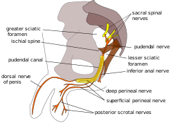 Pudendal nerve and its course through the pudendal canal (labelled in yellow) | |
| Details | |
| Identifiers | |
| Latin | canalis pudendalis |
| TA | A09.5.04.003 |
| FMA | 22071 |
| Anatomical terminology | |
Structure
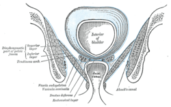
The pudendal canal is formed by the fascia of the obturator internus muscle, or obturator fascia.
It encloses the following:
These vessels and nerve cross the pelvic surface of the obturator internus.
Additional images
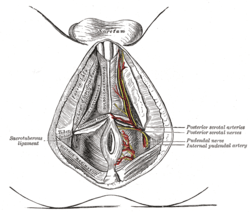 The superficial branches of the internal pudendal artery. (Canal not labeled, but pudendal nerve and internal pudendal artery labeled at bottom right.)
gollark: You have nice stuff like the ability to run `RightCtrl+S` to disable/enable running (in-sandbox) startup thanks to that sandboxing.
gollark: PotatOS applies sandboxing; if it were not for that, OneOS would overwrite potatOS.
gollark: Well, hardly.
gollark: Installing potatOS within OneOS will overwrite OneOS with potatOS; installing OneOS within potatOS will be fine.
gollark: CraftOS is installed by default.
See also
References
This article incorporates text in the public domain from page 421 of the 20th edition of Gray's Anatomy (1918)
External links
- Anatomy image: apmalefrontal4-16 at the College of Medicine at SUNY Upstate Medical University
- Cross section image: pelvis/pelvis-e12-15—Plastination Laboratory at the Medical University of Vienna
- Anatomy photo:41:08-0100 at the SUNY Downstate Medical Center — "The Female Perineum: Contents of the Pudendal Canal"
- Diagram at pudendal.info
- Anatomy image:9087 at the SUNY Downstate Medical Center
- Anatomy image:9448 at the SUNY Downstate Medical Center
| Authority control |
|---|
This article is issued from Wikipedia. The text is licensed under Creative Commons - Attribution - Sharealike. Additional terms may apply for the media files.