Protosteloid
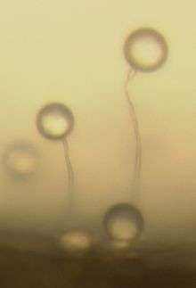
Protosteloid amoebae, also called protostelids, are amoebae that are capable of making simple fruiting bodies consisting of a cellular stalk topped by one or a few spores.[1] All species are microscopic and are typically found on dead plant matter where they consume bacteria, yeasts, and fungal spores. Since protostelids are amoebae that make spores they are considered to be slime molds.[2]

Ecology
Protosteloid amoebae are typically found on dead plant matter, including stems and leaves of herbaceous plants, stems and leaves of grasses, bark of living trees, decaying wood and other types of dead plant matter.[1] Protosteloid amoebae are found on dead plant matter that has fallen on the ground, on dead plant parts that are still hanging in the air and on dead parts of plants that are submerged in a pond.[3][4] They have also been detected on the petals of living flowers and on living tree leaves. Since protosteloid amoebae eat bacteria, yeasts, and fungal spores in the laboratory, it is thought that they also do this in nature.[2] They are thus thought to be predators in the decomposer community.[2]
Distribution
Collections of protostelids have been made from all continents, including the Antarctic peninsula. Protostelids have also been found on isolated islands like Hawaii in the Pacific and Ascension Island in the southern Atlantic,[5][6] indicating that protostelids have a worldwide distribution. Most studies of protostelid distribution have been done in the temperate zones so they are best known from these areas.[7][8] However, tropical studies have turned up protostelids, often in great abundance.[9]
Collection and laboratory culture
Since protosteloid amoebae are microscopic one must bring their substrates, dead plant matter, into the laboratory to find them. Dead plant matter is placed on the agar surface in a petri plate and allowed to incubate for several days to a week. Then the edges of the substrates are scanned with a compound microscope and species are identified by their fruiting body morphology and amoebal morphology.[2]
When protosteloid fruiting bodies are found they can be moved into laboratory culture onto an appropriate food organism or mix of organisms. This is done by picking up fruiting bodies or spores with a sterilised needle and moving them onto agar in a fresh petri plate that has been smeared with a bacterium or yeast upon which the protosteloid amoeba species has been known to grow. If the spores germinate then the protostelid begins eating the food organism and a culture is established.[2]
Taxonomy and relationships
The formal taxonomy of protosteloid amoebae groups them all according to fruiting bodies, mostly leaving out characteristics of the amoebae. Recent studies have shown that all protosteloid amoebae studied to date are probably included in the group known as Amoebozoa or Eumycetozoa.[10][11] However, protosteloid amoebae are not all closely related and some fall within groups of amoebae in which the other amoebae are nonfruiting.[10][11] Therefore, it appears that the ability to make fruiting bodies may have evolved more than once.
References
- Olive, Lindsay S. (1975). The mycetozoans. New York: Academic press. ISBN 9780125262507.
- Spiegel, Frederick W.; Steven L. Stephenson; Harold W. Keller; Donna L Moore; James C. Cavendar (2004). "Mycetozoans". In Gregory M. Mueller; Gerald F. Bills; Mercedes S. Foster (eds.). Biodiversity of fungi : inventory and monitoring methods. New York: Elsevier Academic Press. pp. 547–576. ISBN 0125095511.
- Lindley, Lora A.; Steven L. Stephenson; Frederick W. Spiegel (1 July 2007). "Protostelids and myxomycetes isolated from aquatic habitats". Mycologia. 99 (4): 504–509. doi:10.3852/mycologia.99.4.504. PMID 18065001.
- Spiegel, Frederick W. "A beginner's guide to identifying the protostelids" (PDF). The Eumycetozoan Project. Retrieved 31 May 2012.
- Spiegel, Frederick W.; John D. L. Shadwick; Lora A. Lindley; Matt Brown; Don E. Hemmes (2007). "Protostelids of the Hawaiian archipelago". Innoculum. 59 (2): 38.
- Landolt, John C.; John D.L. Shadwick; Steven L. Stephenson (30 December 2008). "First records of dictyostelids and protostelids from Ascension Island". Sydowia. 60 (2): 235–245.
- Zahn, Geoffrey; Stephenson, Spiegel (11 March 2014). "Ecological distribution of protosteloid amoebae in New Zealand". PeerJ. 2: e296. doi:10.7717/peerj.296. PMC 3961141. PMID 24688872.
- Shadwick, J. D.L.; Stephenson, S. L.; Spiegel, F. W. (2009). "Distribution and ecology of protostelids in Great Smoky Mountains National Park". Mycologia. 101 (3): 320–328. doi:10.3852/08-167. ISSN 0027-5514.
- Moore, Donna L.; Frederick W. Spiegel (July 2000). "Microhabitat Distribution of Protostelids in Tropical Forests of the Caribbean National Forest, Puerto Rico". Mycologia. 92 (4): 616–625. doi:10.2307/3761419.
- Shadwick, Lora L.; FW Spiegel; JDL Shadwick; MW Brown; JD Silberman (August 2009). "Eumycetozoa=Amoebozoa?: SSUrDNA Phylogeny of Protosteloid Slime Molds and Its Significance for the Amoebozoan Supergroup". PLoS ONE. 4 (8): e6754. doi:10.1371/journal.pone.0006754. PMC 2727795. PMID 19707546.
- Lahr, Daniel JG; J Grant; T Nguyen; JH Lin; LA Katz (28 July 2011). "Comprehensive Phylogenetic Reconstruction of Amoebozoa Based on Concatenated Analyses of SSU-rDNA and Actin Genes". PLoS ONE. 6 (7): e22780. doi:10.1371/journal.pone.0022780. PMC 3145751. PMID 21829512.