Parieto-occipital sulcus
The parieto-occipital sulcus (also called the parietooccipital fissure) is a deep fissure in the cerebral cortex that marks the boundary between the cuneus and precuneus, and also between the parietal and occipital lobes. Only a small part can be seen on the lateral surface of the hemisphere, its chief part being on the medial surface.
| Parieto-occipital sulcus | |
|---|---|
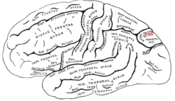 Fig. 726: Lateral surface of left cerebral hemisphere, viewed from the side. | |
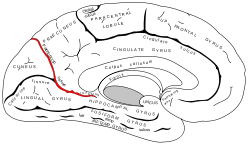 Fig. 727: Medial surface of left cerebral hemisphere. | |
| Details | |
| Identifiers | |
| Latin | sulcus parietooccipitalis, fissura parietooccipitalis |
| NeuroNames | 52 |
| NeuroLex ID | birnlex_1428 |
| TA | A14.1.09.108 |
| FMA | 83754 |
| Anatomical terms of neuroanatomy | |
The lateral part of the parieto-occipital sulcus (Fig. 726) is situated about 5 centimeters (cm) in front of the occipital pole of the hemisphere, and measures about 1.25 cm. in length.
The medial part of the parieto-occipital sulcus (Fig. 727) runs downward and forward as a deep cleft on the medial surface of the hemisphere, and joins the calcarine fissure below and behind the posterior end of the corpus callosum. In most cases it contains a submerged gyrus.
Function
The parieto-occipital lobe has been found in various neuroimaging studies, including PET (positron-emission-tomography) studies,[1][2][3][4] and SPECT (single-photon emission computed tomography) studies,[5][6] to be involved along with the dorsolateral prefrontal cortex during planning.
Gallery
 Animation of left cerebral hemisphere. Parieto-occipital sulcus shown in red.
 Medial surface of right hemisphere. Parieto-occipital sulcus labeled at top right as "*"
 Medial surface of left hemisphere. Parieto-occipital sulcus visible at top left.
References
This article incorporates text in the public domain from page 820 of the 20th edition of Gray's Anatomy (1918)
- Owen, Adrian M.; Doyon, Julien; Petrides, Michael; Evans, Alan C. (1996). "Planning and Spatial Working Memory: a Positron Emission Tomography Study in Humans". European Journal of Neuroscience. 8 (2): 353–364. doi:10.1111/j.1460-9568.1996.tb01219.x.
- Baker, S.C.; Rogers, R.D.; Owen, A.M.; Frith, C.D.; Dolan, R.J.; Frackowiak, R.S.J.; Robbins, T.W. (June 1996). "Neural Systems Engaged by Planning: a PET Study of the Tower of London Task" (PDF). Neuropsychologia. 34 (6): 515–526. doi:10.1016/0028-3932(95)00133-6. hdl:21.11116/0000-0001-A39D-6. PMID 8736565. Retrieved 18 June 2014.
- Dagher, Alain; Owen, Adrian M.; Boecker, Henning; Brooks, David J. (October 1999). "Mapping the Network for Planning: a Correlational PET activation study with the Tower of London Task". Brain. 122 (10): 1973–1987. doi:10.1093/brain/122.10.1973. Retrieved 18 June 2014.
- Rowe, J.B.; Owen, Adrian M.; Johnsrude, Ingrid S.; Passingham, R.E. (2001). "Imaging the Mental Components of a Planning Task" (PDF). Neuropsychologia. 39 (3): 315–327. doi:10.1016/S0028-3932(00)00109-3. Retrieved 18 June 2014.
- Rezai, Karim; Andreasen, Nancy C.; Alliger, Randy; Cohen, Gregg; Swayze, Victor II; O'Leary, Daniel S. (June 1993). "The Neuropsychology of the Prefrontal Cortex". Archives of Neurology. 50 (6): 636–642. doi:10.1001/archneur.1993.00540060066020. Retrieved 18 June 2014.
- Morris, R.G.; Ahmed, S.; Syed, G.M.; Toone, B.K. (December 1993). "Neural Correlates of Planning Ability: Frontal Lobe Activation during the Tower of London Test". Neuropsychologia. 31 (12): 1367–1378. doi:10.1016/0028-3932(93)90104-8. PMID 8127433.
External links
| Wikimedia Commons has media related to Parieto-occipital sulcus. |
- "Anatomy diagram: 13048.000-3". Roche Lexicon - illustrated navigator. Elsevier. Archived from the original on 2012-07-22.
- https://web.archive.org/web/20070316043257/http://www2.umdnj.edu/~neuro/studyaid/Practical2000/Q30.htm
| Authority control |
|---|