Median cubital vein
In human anatomy, the median cubital vein (or median basilic vein) is a superficial vein of the upper limb.[1] It is very clinically relevant as it is routinely used for venipuncture (taking blood) and as a site for an intravenous cannula . It connects the basilic and cephalic vein and becomes prominent when pressure is applied. It lies in the cubital fossa superficial to the bicipital aponeurosis.
| Median cubital vein | |
|---|---|
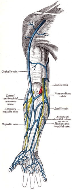 Superficial veins of the upper limb. The median cubital vein is labelled (in Latin) - Vena mediana cubiti. | |
| Details | |
| Source | cephalic vein |
| Drains to | basilic vein |
| Identifiers | |
| Latin | vena mediana cubiti |
| TA | A12.3.08.019 |
| FMA | 22963 |
| Anatomical terminology | |
There exists a fair amount of variation of the median cubital vein. More commonly the vein forms an H-pattern with the cephalic and basilic veins making up the sides. Other forms include an M-pattern, where the vein branches to the cephalic and basilic veins.
Additional images
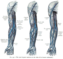 The most frequent variations of the veins of the forearm (schematic).
- Dissection images
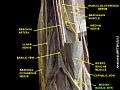 Median basilic vein
gollark: I'm just telling you what the policy says.
gollark: Yes you do.
gollark: You do, although this is irrelevant as it's internal GTech™ policy.
gollark: Exciting news: new policy permits us to convert statements into arbitrary noble gases!
gollark: I was probably not planning to.
See also
References
- Standring, Susan. Gray's anatomy: the anatomical basis of clinical practice (41 ed.). Elsevier Limited. pp. 837–861. ISBN 978-0-7020-5230-9.
External links
- Anatomy photo:07:st-0703 at the SUNY Downstate Medical Center
- Radiology image: UpperLimb:18VenoFo from Radiology Atlas at SUNY Downstate Medical Center (need to enable Java)
| Authority control |
|---|
This article is issued from Wikipedia. The text is licensed under Creative Commons - Attribution - Sharealike. Additional terms may apply for the media files.