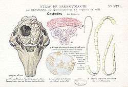
Coenurosis
Coenurosis, also known as caenurosis, coenuriasis, gid or sturdy, is a parasitic infection that develops in the intermediate hosts of some tapeworm species (Taenia multiceps,[1] T. serialis,[2] T. brauni, or T. glomerata). It is caused by the coenurus, the larval stage of these tapeworms. The disease occurs mainly in sheep and other ungulates,[3] but it can also occur in humans by accidental ingestion of tapeworm eggs.

Adult worms of these species develop in the small intestine of the definitive hosts (dogs, foxes and other canids), causing a disease from the group of taeniasis.[4] Humans cannot be definitive hosts for these species of tapeworms.
History
Coenurosis is a rare disease in humans that was not diagnosed until the twentieth century. The four Taenia spp. that cause coenurosis were first diagnosed as follows:
- Taenia multiceps: In 1913, a man in Paris passed away after suffering from multiple neurological symptoms. During his autopsy, two coenuri were found in his brain.[5]
- Taenia glomerate: In 1919, the first coenurosis case due to T. glomerate was diagnosed in Nigeria.[6]
- Taenia serialis: In 1933, the first coenurosis case due to T. serialis was diagnosed in France. [5]
- Taenia brauni: In 1956, the first coenurosis case due to T. brauni was diagnosed in Africa. T. brauni is mainly associated with the coenurosis cases in Africa.[6]
Life cycle
The eggs of T.multiceps, T. glomerate, T. serialis, and T. brauni are shed in the feces of infected hosts into the environment.[7] The eggs are then ingested by an intermediate host, where the eggs hatch in intestines and release oncospheres.[7] Oncospheres are the larval form of tapeworms that contain hooks for attaching to the host’s tissues.[8] The oncospheres continue to move through the bloodstream of the intermediate host until they find suitable organs to inhabit.[8] The oncospheres can bind to the eyes, the brain, skeletal muscle, and subcutaneous tissue. Once the oncospheres reach their destination, they take about three months to develop into coneuri.[7] Coenuri are white, fluid filled structures that are 3-10 centimeters in diameter.[9] Coenuri have a collapsed membrane and several protoscolices on the interior.[9] The coenuri cysts that inhabit the central nervous system have multiple cavities, and the coenuri cysts that are not in the central nerve system have one cavity.[9] The disease is transferred to the definitive host when the host digests the tissue of the intermediate host. Next, eggs hatch in the intestine of the definitive host and circulate in the bloodstream until they reach suitable organs.[7]
Symptoms and diagnosis
The symptoms for coenurosis vary depending on where the cyst is located. When the cyst is in the spinal canal, it causes inflammation of the membranes that surround the spinal cord.[10] This condition is called arachnoiditis. When the cyst is in the eye, it causes decreased vision and leads to blindness.[10] When the cyst is in muscular or subcutaneous issue, it causes painful lesions to form. When the cyst is in the brain, the patient will experience neurological symptoms.[10] These symptoms include headaches, seizures, ataxia, vomiting, monoplegia, and hemiplegia. Since coenurosis is very rare in humans, there are not many ways to diagnose the disease.[10] The most common method for diagnosis involves using CT or MRI scans to visualize the cysts in the body.[6] Some other clinical findings that can be used for diagnosis include papilledemas, hypoglycorrhacia, and high intracranial pressure caused by obstruction of the ventricles.[6]
Prevention and treatment
Coenurosis is a rare disease in humans, so no vaccine has been created yet.[5] The best way to practice prevention would be to avoid eating food and vegetables that are grown in fields fertilized with dog manure.[5] Also, if farmers used methods other than manure to fertilize their fields, it would help prevent the spread of coenurosis.[5] The main method for treating the disease would be the surgical removal of cysts. Alternative methods for treating coenurosis include administering anthelmintics and glucocorticoids to the patients.[5] Praziquantel, Niclosamide, and Albendazole are the anthelminthics that are commonly used to treat coenurosis. Praziquantel works by causing contraction of the parasite by altering the permeability of the cell membrane.[11] This causes the parasite to lose its intracellular calcium. The drug also causes vacuolization and disintegration of the parasite.[11] Niclosamide works by uncoupling oxidative phosphorylation and inhibiting glucose uptake in the parasite.[12] Albendazole works by binding to β- tubulin sites and inhibiting their polymerization into microtubules.[13] This decreases the absorption ability of the parasite, leading to decreased glucose uptake and glycogen stores.[13] The lack of glucose prevents the parasite from making enough ATP leading to the parasite’s death. Dexamethasone is the glucocorticoid that is normally administered for coneurosis.[5] It inhibits pro-inflammatory signals and promotes anti-inflammatory signals in the body.[14] Dexamethasone also causes decreased vasodilation in capillaries and inhibits apoptosis in neutrophils.[14]
Epidemiology
Since the incidence of coenurosis is so low in humans, there are no endemic areas. Most cases occur in Europe and Africa and there are few cases in the Western Hemisphere.[9]
Hosts
The definitive hosts for coenurosis are dogs, foxes, and other canids.[15] The intermediate hosts for coenurosis can vary depending on the Taenia spp. In T. multiceps, sheep are the intermediate hosts, but goats, cattle, horses, and antelopes are also common hosts.[15] T. multiceps can affect any tissue, but it normally targets the brain in animal hosts. In T. serialis, rabbits and rodents are the intermediate hosts.[15] T. serialis commonly targets subcutaneous and intramuscular tissue. In T. brauni and T. glomerata, gerbils are the intermediate host. T. brauni and T. glomerate larvae tend to inhabit the muscles. Intermediate hosts can be infected with either chronic or acute coenurosis.[15] Chronic coenurosis is the more common form, and it occurs primarily in young sheep.
In wild animals
Although coenurosis is more commonly associated with domestic animals, it has also been documented in wildlife, such as in mountain ungulates in the French Alps. It is believed that the ungulates are being contaminated by infected sheep. Understanding how this disease is transmitted from sheep to wild animals is important in managing the spread of this potentially dangerous zoonotic disease. A potential management strategy would be for farmers to dispose of animal carcasses found on their land. In Ethiopia, gelada monkeys with coenurosis were found to affect the fitness of other primates.[16][17] Animals infected with the disease tend to hide from predators and might not be seen by humans.
See also
References
- University of Pennsylvania - Veterinary Medicine: Taenia multiceps Homepage Archived 2010-07-10 at the Wayback Machine
- University of Pennsylvania - Veterinary medicine: Taenia serialis Homepage Archived 2010-07-10 at the Wayback Machine
- Stanford University: Coenurosis - Hosts
- Stanford University: Taeniasis
- "Coenurosis". web.stanford.edu. Retrieved 2020-04-30.
- Hermos, John A.; Healy, George R.; Schultz, Myron G.; Barlow, John; Church, William G. (1970-08-31). "Fatal Human Cerebral Coenurosis". JAMA. 213 (9): 1461–1464. doi:10.1001/jama.1970.03170350029006. ISSN 0098-7484.
- Armon, Robert; Cheruti, Uta (2012). "Environmental Aspects of Zoonotic Diseases". Water Intelligence Online. 11. doi:10.2166/9781780400761. ISSN 1476-1777.
- Smyth, James Desmond (2007). The physiology and biochemistry of cestodes. Cambridge University Press. ISBN 978-0-521-03895-9. OCLC 836624725.
- Ryan, Edward T.; Hill, David Russell; Solomon, Tom; Endy, Timothy P.; Aronson, Naomi, eds. (25 March 2019). Hunter's tropical medicine and emerging infectious diseases. ISBN 978-0-323-62550-0. OCLC 1096243611.
- Principles and Practice of Pediatric Infectious Diseases. 2018. doi:10.1016/c2013-0-19020-4. ISBN 9780323401814.
- Chai, Jong-Yil (2013). "Praziquantel Treatment in Trematode and Cestode Infections: An Update". Infection & Chemotherapy. 45 (1): 32. doi:10.3947/ic.2013.45.1.32. ISSN 2093-2340. PMID 24265948.
- Chen, Wei; Mook, Robert A.; Premont, Richard T.; Wang, Jiangbo (January 2018). "Niclosamide: Beyond an antihelminthic drug". Cellular Signalling. 41: 89–96. doi:10.1016/j.cellsig.2017.04.001. ISSN 0898-6568. PMC 5628105. PMID 28389414.
- Firth, Mary (1984). Albendazole in helminthiasis. Royal Society of Medicine. ISBN 978-0-19-922001-4. OCLC 10207175.
- Tsurufuji, Susumu (1981). "Molecular mechanisms and mode of action of anti-inflammatory steroids". Mechanisms of Steroid Action. Biological Council The Co-ordinating Committee for Symposia on Drug Action. Macmillan Education UK. pp. 85–95. doi:10.1007/978-1-349-81345-2_7. ISBN 978-1-349-81347-6.
- Rahsan, Yilmaz; Nihat, Yumusak; Bestami, Yilmaz; Adnan, Ayan; Nuran, Aysul (2018-03-30). "Histopathological, immunohistochemical, and parasitological studies on pathogenesis of Coenurus cerebralis in sheep". Journal of Veterinary Research. 62 (1): 35–41. doi:10.2478/jvetres-2018-0005. ISSN 2450-8608.
- Schneider-Crease I, Griffin RH, Gomery MA, Bergman TJ, Beehner JC (2017). "High mortality associated with tapeworm parasitism in geladas (Theropithecus gelada) in the Simien Mountains National Park, Ethiopia". American Journal of Primatology. 79 (9): e22684. doi:10.1002/ajp.22684. PMID 28783206.
- Nguyen N, Fashing PJ, Boyd DA, Barry TS, Burke RJ, Goodale CB, Jones SC, Kerby JT, Kellogg BS, Lee LM, Miller CM, Nurmi NO, Ramsay MS, Reynolds JD, Stewart KM, Turner TJ, Venkataraman VV, Knauf Y, Roos C, Knauf S (2017). "Fitness impacts of tapeworm parasitism on wild gelada monkeys at Guassa, Ethiopia". American Journal of Primatology. 77 (5): 579–594. doi:10.1002/ajp.22379. PMID 25716944.
External links
- Stanford University: Coenurosis