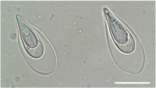
Thelohanellus kitauei
T. kitauei is a myxozoan endoparasite identified as the agent of intestinal giant-cystic disease (IGCD) of common carp Cyprinus carpio. The species was first identified in Japan, in 1980[1] and later formally described by Egusa & Nakajima.[2] Fan[3] subsequently reported the parasite in China, and several other reports from carp and Koi carp in China and Korea followed.[4][5][6] Reports referred to an intestinal infection, swelling and emaciation of fish due to blockage of the intestinal tract by giant cysts. The intestine of carp was believed to be the only infection site of T. kitauei until Zhai et al.[7] reported large cysts of T. kitauei in the skin, with morphologically similar and molecularly identical spores. T. kitauei has been recognized as the most detrimental disease of farmed carp in Asia with around 20% of farmed carp killed annually.[8] In 2014, the genome of T. kitauei was sequenced,[8] and in 2016, its life cycle was found to include the oligochaete Branchiura sowerbyi.[9] Infected oligochaete worms were first discovered in Hungary and raised concerns of the introduction of T. kitauei into European carp culture ponds, since it was believed to be endemic to Asia. However, the related disease (IGCD) has not yet been reported in Europe.

Taxonomy
Valid taxonomic name: Thelohanellus kitauei Egusa & Nakajima, 1981.
Junior synonym: Thelohanellus xinyangensis Xie, Gong, Xiao, Guo, Li et Guo, 2000.
Life cycle
The life cycle of T. kitauei involves two hosts. Branchiura sowerbyi (Oligochaeta, Naididae, Branchiurinae) is the definitive host, in which actinosporean spores of the aurantiactinomyxon type are formed and released from the intestinal tract of the worms. These actinospores then infect the intermediate host, common carp, and after parasite multiplication, myxosporean spores are formed in the carp intestine. Myxospores are then released with the faeces of the fish host or after its death, and taken up by B. sowerbyi while feeding on the sediment.
Pathology and clinical signs
The intestines of diseased carp develop large cysts containing spores of T. kitauei. The cyst size ranges from 2 cm to 3.6 cm in diameter. Histopathology indicates that T. kitauei first invades the submucosa of the host intestine and then moves into the mucosa layers where spores are formed, with spores entering the body cavity of the hosts after disruption of mucosa layers.[10] Affected fish stocks can suffer from mortalities reaching 100%. Fish die from starvation as nutrient uptake and passage of food through infected intestines is limited. Infections in the skin cause large, irregular tumour-like lesions bearing cysts with spores. These cause exfoliation of epidermis and the stratum spongiosum.
Impact
In 2010, in China, IGCD led to economic losses of approximately US$50 Million.[8]
Diagnosis
Intestinal cysts are large and easily visible to the naked eye (Fig. 2), cysts on the skin are also large. Myxospores within the cysts should match the descriptions of Egusa & Nakajima, 1981[2] or Zhai et al. 2016.[7] They are egg-shaped balloon-like sacks, 33.4 μm by 15.0 μm in size on average. 18S rDNA sequences are available on Genbank under the accession numbers KU664644, JQ690367, HM624024, MH329616, KR872638, KU664643, GQ396677, MF536693; genome data is available under the Bioproject accession number PRJNA193083. A qPCR detection assay was developed by Seo et al.,[11] but its specificity requires confirmation as closely related species show only minor 18S rDNA sequence divergence.
Treatments
Presently, there are no treatments against myxozoans in fish destined for human consumption.
Other control strategies
No other control strategies have been identified.
Research
Most research on this parasite is performed in Japan, China and Korea, where IGCD affects a large number of carp stocks. Within the EU-funded Horizon 2020 Project ParaFishControl the invertebrate host of T. kitauei was elucidated in Hungary, though this country is thought to be an IGCD-free carp culture area. Ongoing studies within this project investigate the presence of T. kitauei in environmental samples in carp culture ponds in different countries in Europe and focus on detecting a potentially different infection site in fish, as large scale screening of intestines did not reveal T. kitauei infections, even at sites where the parasite was detected in environmental samples.
References
- Kitaue K (1980) Intestinal giant-cystic disease affecting the carp, caused by Thelohanellus sp. Fish Pathology, 14, 145-146
- Egusa S, Nakajima K (1981) A new Myxozoa Thelohanellus kitauei, the cause of intestinal giant cystic disease of carp. Fish Pathology 15, 213-218
- Fan ZG (1985) Study of thelohanellosis from common carp. Freshwater Fish 5, 16-18
- Liu Y, Whipps CM, Liu WS, Zeng LB, Gu ZM (2011) Supplemental diagnosis of a myxozoan parasite from common carp Cyprinus carpio: synonymy of Thelohanellus xinyangensis with Thelohanellus kitauei. Veterinary Parasitology 178, 355-359
- Shin SP, Jee H, Han JE, Kim JH, Choresca CH, Jun JW, Kim DY, Park SC (2011) Surgical removal of an anal cyst caused by a protozoan parasite (Thelohanellus kitauei) from a koi (Cyprinus carpio) Journal of the American Veterinary Medicine Association 238, 784-786.
- Shin SP, Kim JH, Choresca CH, Han JE, Jun JW, Park SC (2013) Molecular identification and phylogenetic characterisation of Thelohanellus kitauei Acta Veterinaria Hungarica 61, 30-35.
- Zhai Y, Gu Z, Guo Q, Wu Z, Wang H, Liu Y (2016) New type of pathogenicity of Thelohanellus kitauei Egusa & Nakajima, 1981 infecting the skin of common carp Cyprinus carpio L. Parasitology International 65, 78-82.
- YanYL, Xiong J, Zhou ZG, Huo FM, Miao W, Ran C et al. (2014) The genome of the myxosporean Thelohanellus kitauei shows adaptations to nutrient acquisition within its fish host. Genome Biology and Evolution 6, 3182-3198.
- Zhao D, Borkhanuddin MH, Wang W, Liu Y, Cech G, Zhai Y, Székely C (2016) The life cycle of Thelohanellus kitauei(Myxozoa: Myxosporea) infecting common carp (Cyprinus carpio) involves aurantiactinomyxon in Branchiura sowerbyi. Parasitology Research 115, 4317-4325.
- Ye L, LU M, Quan K, Li W, Zou H, Wu S, Wang J, Wang G (2016) Intestinal disease of scattered mirror carp Cyprinus carpio caused by Thelohanellus kitauei and notes on the morphology and phylogeny of the myxosporean from Sichuan Province, southwest China. Chinese Journal of Oceanology and Limnology 35, 587-596.
- Seo JS, Jeon EJ, Kim MS, Woo SH, Kim JD, Jung SH, Park MA, Jee BY, Kim JW, Kim YC, Lee EH (2012) Molecular identification and real-time quantitative PCR (qPCR) for rapid detection of Thelohanellus kitauei, a myxozoan parasite causing Intestinal Giant Cystic Disease in the Israel Carp. Korean Journal of Parasitology 50, 103-111.