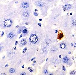
TUNEL assay
Terminal deoxynucleotidyl transferase dUTP nick end labeling (TUNEL) is a method for detecting DNA fragmentation by labeling the 3′- hydroxyl termini in the double-strand DNA breaks generated during apoptosis.[1][2]

Method
TUNEL is a method for detecting apoptotic DNA fragmentation, widely used to identify and quantify apoptotic cells, or to detect excessive DNA breakage in individual cells.[3] The assay relies on the use of terminal deoxynucleotidyl transferase (TdT), an enzyme that catalyzes attachment of deoxynucleotides, tagged with a fluorochrome or another marker, to 3'-hydroxyl termini of DNA double strand breaks. It may also label cells having DNA damaged by other means than in the course of apoptosis.
History
The fluorochrome-based TUNEL assay applicable for flow cytometry, combining the detection of DNA strand breaks with respect to the cell cycle-phase position, was originally developed by Gorczyca et al.[4] Concurrently, the avidin-peroxidase labeling assay applicable for light absorption microscope was described by Gavrieli et al.[5] Since 1992 the TUNEL has become one of the main methods for detecting apoptotic programmed cell death.[6] However, for years there has been a debate about its accuracy, due to problems in the original assay which caused necrotic cells to be inappropriately labeled as apoptotic.[7] The method has subsequently been improved dramatically and if performed correctly should only identify cells in the last phase of apoptosis.[8][9] New methods incorporate the dUTPs modified by fluorophores or haptens, including biotin or bromine, which can be detected directly in the case of a fluorescently-modified nucleotide (i.e., fluorescein-dUTP), or indirectly with streptavidin or antibodies, if biotin-dUTP or BrdUTP are used, respectively. The most sensitive of them is the method utlilizing incorporation of BrdUTP by TdT followed by immunocytochemical detection of BrdU.[10]
References
- Gorczyca W, Traganos F, Jesionowska H, Darzynkiewicz Z (1993). "Presence of DNA strand breaks and increased sensitivity of DNA in situ to denaturation in abnormal human sperm cells. Analogy to apoptosis of somatic cells". Exp Cell Res. 207 (1): 202–205. doi:10.1006/excr.1993.1182. PMID 8391465.
- Lozano GM, Bejarano I, Espino J, González D, Ortiz A, García JF, Rodríguez AB, Pariente JA (2009). "Density gradient capacitation is the most suitable method to improve fertilization and to reduce DNA fragmentation positive spermatozoa of infertile men". Anatolian Journal of Obstetrics & Gynecology. 3 (1): 1–7.
- Lozano GM, Bejarano I, Espino J, González D, Ortiz A, García JF, Rodríguez AB, Pariente JA (2009). "Relationship between Caspase Activity and Apoptotic Markers in Human Sperm in Response to Hydrogen Peroxide and Progesterone". Journal of Reproduction and Development. 55 (6): 615–621. doi:10.1262/jrd.20250. PMID 19734695.
- Gorczyca W, Bruno S, Darzynkiewicz RJ, Gong J, Darzynkiewicz Z (1992). "DNA strand breaks occurring during apoptosis: Their early in situ detection by the terminal deoxynucleotidyl transferase and nick translation assays and prevention by serine protease inhibitors". International Journal of Oncology. 1 (6): 639–648. doi:10.3892/ijo.1.6.639. PMID 21584593.
- Gavrieli Y, Sherman Y, Ben-Sasson SA (1992). "Identification of programmed cell death in situ via specific labeling of nuclear DNA fragmentation". J. Cell Biol. 119 (3): 493–501. doi:10.1083/jcb.119.3.493. PMC 2289665. PMID 1400587.CS1 maint: multiple names: authors list (link)
- Darzynkiewicz Z, Galkowski D, Zhao H (2008). "Analysis of apoptosis by cytometry using TUNEL assay". Methods. 44 (3): 250–254. doi:10.1016/j.ymeth.2007.11.008. PMC 2295206. PMID 18314056.
- Grasl-Kraupp B, Ruttkay-Nedecky B, Koudelka H, Bukowska K, Bursch W, Schulte-Hermann R (1995). "In situ detection of fragmented DNA (TUNEL assay) fails to discriminate among apoptosis, necrosis, and autolytic cell death: a cautionary note". Hepatology. 21 (5): 1465–8. doi:10.1002/hep.1840210534. PMID 7737654.
- Negoescu A, Lorimier P, Labat-Moleur F, Drouet C, Robert C, Guillermet C, Brambilla C, Brambilla E (1996). "In situ apoptotic cell labeling by the TUNEL method: improvement and evaluation on cell preparations". J Histochem Cytochem. 44 (9): 959–68. doi:10.1177/44.9.8773561. PMID 8773561.
- Negoescu A, Guillermet C, Lorimier P, Brambilla E, Labat-Moleur F (1998). "Importance of DNA fragmentation in apoptosis with regard to TUNEL specificity". Biomed Pharmacother. 52 (6): 252–8. doi:10.1016/S0753-3322(98)80010-3. PMID 9755824.
- Li X, Darzynkiewicz Z (1995). "Labeling DNA strand breaks with BrdUTP. Detection of apoptosis and cell proliferation". Cell Proliferation. 28 (11): 571–579. doi:10.1111/j.1365-2184.1995.tb00045.x. PMID 8555370.
External links
- TUNEL at the US National Library of Medicine Medical Subject Headings (MeSH)