Synthesis of nanoparticles by fungi
Throughout human history, fungi have been utilized as a source of food and harnessed to ferment and preserve foods and beverages. In the 20th century, humans have learned to harness fungi to protect human health (antibiotics, anti-cholesterol statins, and immunosuppressive agents), while industry has utilized fungi for large scale production of enzymes, acids, and biosurfactants.[1] With the advent of modern nanotechnology in the 1980s, fungi have remained important by providing a greener alternative to chemically synthesized nanoparticle.[2]
Background

A nanoparticle is defined as having one dimension 100 nm or less in size. Environmentally toxic or biologically hazardous reducing agents are typically involved in the chemical synthesis of nanoparticles[2] so there has been a search for greener production alternatives.[3][4] Current research has shown that microorganisms, plant extracts, and fungi can produce nanoparticles through biological pathways.[2][3][5] The most common nanoparticles synthesized by fungi are silver and gold, however fungi have been utilized in the synthesis other types of nanoparticles including zinc oxide, platinum, magnetite, zirconia, silica, titanium, and cadmium sulfide and cadmium selenide quantum dots.
Silver nanoparticle production
Synthesis of silver nanoparticles has been investigated utilizing many ubiquitous fungal species including Trichoderma,[6][7] Fusarium,[8] Penicillium,[9] Rhizoctonia, Pleurotus and Aspergillus.[10] Extracellular systhesis has been demonstrated by Trichoderma virde, T. reesei, Fusarium oxysporm, F. semitectum, F. solani, Aspergillus niger, A. flavus,[11] A. fumigatus, A. clavatus, Pleurotus ostreatus, Cladosporium cladosporioides,[6] Penicillium brevicompactum, P. fellutanum, an endophytic Rhizoctonia sp., Epicoccum nigrum, Chrysosporium tropicum, and Phoma glomerata, while intracellular synthesis was shown to occur in a Verticillium [12] species, and in Neurospora crassa.
Gold nanoparticle production
Synthesis of gold nanoparticles has been investigated utilizing Fusarium,[13] Neurospora,[14] Verticillium, yeasts,[15][16] and Aspergillus. Extracellular gold nanoparticle synthesis was demonstrated by Fusarium oxysporum, Aspergillus niger, and cytosolic extracts from Candida albican. Intracellular gold nanoparticle synthesis has been demonstrated by a Verticillum species, V. luteoalbum,[17]
Miscellaneous nanoparticle production
In addition to gold and silver, Fusarium oxysporum has been used to synthesize zirconia, titanium, cadmium sulfide and cadmium selenide nanosize particles. Cadmium sulfide nanoparticles have also been synthesized by Trametes versicolor, Schizosaccharomyces pombe, and Candida glabrata.[18] The white-rot fungus Phanerochaete chrysosporium has also been demonstrated to be able to synthesize elemental selenium nanoparticles.[19]
Culture techniques and conditions
Culture techniques and media vary depending upon the requirements of the fungal isolate involved, however the general procedure consist of the following: fungal hyphae are typically placed in liquid growth media and placed in shake culture until the fungal culture has increased in biomass. The fungal hyphae are removed from the growth media, washed with distilled water to remove the growth media, placed in distilled water and incubated on shake culture for 24 to 48 hours. The fungal hyphae are separated from the supernatant, and an aliquot of the supernatant is added to 1.0 mM ion solution. The ion solution is then monitored for 2 to 3 days for the formation of nanoparticles. Another common culture technique is to add washed fungal hyphae directly into 1.0 mM ion solution instead of utilizing the fungal filtrate. Silver nitrate is the most widely used source of silver ions, but silver sulfate has also been utilized. Choloroauric acid is generally used as the source of gold ions at various concentrations (1.0 mM[13] and 250 mg to 500 mg[17] of Au per liter). Cadmium sulfide nanoparticle synthesis for F. oxysporum was conducted using a 1:1 ratio of Cd2+ and SO42− at a 1 mM concentration.[20] Gold nanoparticles can vary in shape and size depending on the pH of the ion solution.[17] Gericke and Pinches (2006) reported that for V. luteoalbum small (cc.10 nm) spherical gold nanoparticles are formed at pH 3, larger (spherical, triangular, hexagon and rods) gold nanoparticles are formed at pH 5, and at pH 7 to pH 9 the large nanoparticles tend to lack a defined shape. Temperature interactions for both silver and gold nanoparticles were similar; a lower temperature resulted in larger nanoparticles while higher temperatures produced smaller nanoparticles.[17]
Analytical techniques
Visual observations
For externally synthesized silver nanoparticles the silver ion solution generally becomes brownish in color,[7][8][9] but this browning reaction may be absent. For fungi that synthesize intracellular silver nanoparticles, the hyphae darken to a brownish color while the solution remains clear. In both cases the browning reaction is attributed to the surface plasmon resonance of the metallic nanoparticles.[6][21] For external gold nanoparticle production, the solution color can vary depending on the size of the gold nanoparticles; smaller particles appear pink while large particles appear purple. Intracellular gold nanoparticle synthesis typically turns the hyphae purple while the solution remains clear. Externally synthesized cadmium sulfide nanoparticles were reported to make the solution color appear bright yellow.[20]
Analytical tools
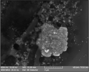
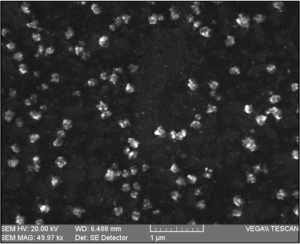
Scanning electron microscopy (SEM), transmission electron microscopy (TEM), energy dispersive analysis of X-ray (EDX), UV-vis spectroscopy, and X-ray diffraction are used to characterize different aspects of nanoparticles. Both SEM and TEM can be used to visualize the location, size, and morphology of the nanoparticles, while UV-vis spectroscopy can be used to confirm the metallic nature, size and aggregation level. Energy dispersive analysis of X-ray is used to determine elemental composition, and X-ray diffraction is used to determine chemical composition and crystallographic structure. UV-Vis absorption peaks for silver, gold, and cadmium sulfide nanoparticles can vary depending on particle size: 25-50 nm silver particles peak ca. 415 nm, gold nanoparticles 30-40 nm peak ca. 450 nm, while a cadmium sulfide absorption edge ca. 450 is indicative of quantum size particles.[20] Larger nanoparticle of each type will have UV-Vis absorption peaks or edges that shift to longer wavelengths while smaller nanoparticles will have UV-Vis absorption peaks or edges that shift to shorter wavelengths.
Formation mechanisms
Gold and silver

Nitrate reductase was suggested to initiate nanoparticle formation by many fungi including Penicillium species, while several enzymes, α-NADPH-dependent reductases, nitrate-dependent reductases and an extracellular shuttle quinone, were implicated in silver nanoparticle synthesis for Fusarium oxysporum. Jain et al. (2011) indicated that silver nanoparticle synthesis for A. flavus occurs initially by a "33kDa" protein followed by a protein (cystein and free amine groups) electrostatic attraction which stabilizes the nanoparticle by forming a capping agent.[11] Intracellular silver and gold nanoparticle synthesis is not fully understood but similar fungal cell wall surface electrostatic attraction, reduction, and accumulation has been proposed.[20] External gold nanoparticle synthesis by P. chrysosporium was attributed to laccase, while intracellular gold nanoparticle synthesis was attributed to ligninase.[20]
Cadmium sulfide
Cadmium sulfide nanoparticle synthesis by yeast involves sequestration of Cd2+ by glutathione-related peptides followed by reduction within the cell. Ahmad et al. (2002) reported that cadmium sulfide nanoparticle synthesis by Fusarium oxysporum was based on a sulfate reductase (enzyme) process.
References
- Barredo JL, ed. (2005). "Microbial cells and enzymes". Microbial enzymes and biotransformations. pp. 1–10. ISBN 978-1-58829-253-7.
- Ghorbani, HR; Safekordi AA; Attar H; Rezayat Sorkhabadi SM (2011). "Biological and non-biological methods for silver nanoparticles synthesis". Chemical and Biochemical Engineering Quarterly. 25: 317–326.
- Abou El-Nour, MM; Eftaiha A; Al-Warthan A; Ammar RAA (2010). "Synthesis and application of silver nanoparticles". Arabian Journal of Chemistry. 3 (3): 135–140. doi:10.1016/j.arabjc.2010.04.008.
- Popescu, M; Velea A; Lőrinczi A (2010). "Biogenic production of nanoparticles". Digest J of Nanomaterials and Biostructures. 5: 1035–1040.
- Sastry, M; Ahmad A; Khan MI; Kumar R (2003). "Biosynthesis of metal nanoparticles using fungi and actinomycete". Current Science. 85: 162–170.
- Vahabi, K; Mansoori GA; Karimi S (2011). "Biosynthesis of silver nanoparticles by fungus Trichoderma reesei: a route for large-scale production of AgNPs". Insciences Journal. 1: 65–79. doi:10.5640/insc.010165.
- Basavaraja, S; Balaji SD; Lagashetty A; Rajasab AH; Venkataraman A (2008). "Extracellular biosynthesis of silver nanoparticles using the fungus Fusarium semitectum". Materials Research Bulletin. 45 (5): 1164–1170. doi:10.1016/j.materresbull.2007.06.020.
- Durán, N; Marcato PD; Alves OL; IH de Souza G; Esposito E (2005). "Mechanistic aspects of biosynthesis of silver nanoparticles by several Fusarium oxysporum strains". Journal of Nanotechnology. 3: 8. doi:10.1186/1477-3155-3-8. PMC 1180851. PMID 16014167.
- Naveen, H; Kumar G; Karthik L; Roa B (2010). "Extracellular biosynthesis of silver nanoparticles using the filamentous fungus Penicillium sp". Archives of Applied Science Research. 2: 161–167.
- Bhainsa, KC; D’Sousa SF (2006). "Extracellular biosynthesis of silver nanoparticles using the fungus Aspergillus fumigatas". Colloids and Surfaces B: Biointerfaces. 47 (2): 160–164. doi:10.1016/j.colsurfb.2005.11.026. PMID 16420977.
- Jain, N; Jain, N., Bhargava A, Majumdar S, Tarafdar J, Panwar J; Majumdar, Sonali; Tarafdar, J. C.; Panwar, Jitendra (2011). "Extracellular biosynthesis and characterization of silver nanoparticles using Aspergillus flavus NJP08: a mechanism perspective". Nanoscale. 3 (2): 635–641. Bibcode:2011Nanos...3..635J. doi:10.1039/c0nr00656d. PMID 21088776.CS1 maint: multiple names: authors list (link)
- Mukherjee, P; Ahmad A, Mandal D, Senapati S, Sainkar S, Khan M, Parishcha R, Ajaykumar P, Alam M, Kumar R, Sastry M; Mandal, Deendayal; Senapati, Satyajyoti; Sainkar, Sudhakar R.; Khan, Mohammad I.; Parishcha, Renu; Ajaykumar, P. V.; Alam, Mansoor; Kumar, Rajiv; Sastry, Murali (2001). "Fungus-mediated synthesis of silver nanoparticles and their immobilization in the mycelial matrix; a novel biological approach to nanoparticle synthesis". Nano Letters. 1 (10): 515–519. Bibcode:2001NanoL...1..515M. doi:10.1021/nl0155274.CS1 maint: multiple names: authors list (link)
- Mukherjee, P; Senapati S; Mandal D; Ahmad A; Khan M; Kumar R; Sastry M (2002). "Extracellular synthesis of gold nanoparticles by the fungus Fusarium oxysporum". ChemBioChem. 3 (5): 461–463. doi:10.1002/1439-7633(20020503)3:5<461::AID-CBIC461>3.0.CO;2-X. PMID 12007181.
- Castro-Longoria, E; Vilchis-Nestor A,and Avales-Borja M; Avalos-Borja, M. (2011). "Biosynthesis of silver, gold, and bimetallic nanoparticles using the filamentous fungus Neurospora crassa". Colloids and Surface B:Biointerfaces. 83: 42–48. doi:10.1016/j.colsurfb.2010.10.035. PMID 21087843.
- Agnihotri, M; Joshi S; Kumar A; Zinjarde S; Kulkarni S (2009). "Biosynthesis of gold nanoparticles by the tropical marine yeast Yarrowia lipolytica NCIM 3589". Material Letters. 63 (15): 1231–1234. doi:10.1016/j.matlet.2009.02.042.
- Chauhan, A; Zubair S; Tufail S; Sherwani A; Sajid M; Raman S; Azam A; Owais M (2011). "Fungus-mediated biological synthesis of gold nanoparticles: potential in detection of liver cancer". International Journal of Nanomedicine. 6: 2305–2319. doi:10.2147/ijn.s23195. PMC 3205127. PMID 22072868.
- Gericke, M; Pinches A (2006). "Biological synthesis of metal nanoparticles". Hydrometallurgy. 83 (1–4): 132–140. doi:10.1016/j.hydromet.2006.03.019.
- Li, X; Xu H; Chen Z; Chen G (2011). "Biosynthesis of nanoparticles by microorganisms and their applications". Journal of Nanomaterials. 2011: 1–16. doi:10.1155/2011/270974.
- Espinosa-Ortiz, EJ; Gonzalez-Gil G; Saikaly PE; van Hullebusch ED; Lens PNL (2014). "Effects of selenium oxyanions on the white-rot fungus Phanerochaete chrysosporium". Appl Microbiol Biotechnol. 99 (5): 2405–2418. doi:10.1007/s00253-014-6127-3.
- Ahmad, A; Mukherjee P; Mandal D; Senapati S; Khan M; Kumar R; Sastry M (2002). "Enzyme mediated extracellular synthesis of CdS nanoparticles by the fungus, Fusarium oxysporum". Journal of the American Chemical Society. 124 (41): 12108–12109. doi:10.1021/ja027296o.
- Shankar, S; Ahmad A; Sastry M (2003). "Geranium leaf assisted biosynthesis of silver nanoparticles". Biotechnol. Prog. 19 (6): 1627–1631. doi:10.1021/bp034070w. PMID 14656132.