Scleral spur
The scleral spur is an annular structure composed of collagen in the human eye, a protrusion of the sclera into the anterior chamber. It is the origin of the longitudinal and circular fibres (which swerve acutely from the spur to run circumferentially as a sphincter near the periphery of the lens)[1] of the ciliary muscle and is attached anteriorly to the trabecular meshwork.
| Scleral spur | |
|---|---|
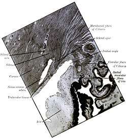 Enlarged general view of the iridial angle (Scleral spur appear at upper right) | |
| Details | |
| System | Visual system |
| Identifiers | |
| Latin | Calcar sclerae |
| Anatomical terms of neuroanatomy | |
Role in treatment of Glaucoma
Open-angle glaucoma (OAG) and closed-angle glaucoma (CAG) may be treated by muscarinic receptor agonists (e.g., pilocarpine), which cause rapid miosis and contraction of the ciliary muscles, this pulls the scleral spur and results in the trabecular meshwork being stretched and separated. This opens the fluid pathways and facilitates drainage of the aqueous humour into the canal of Schlemm and ultimately decreasing intraocular pressure.[2]
References
- Gray's Anatomy: The Anatomical Basis of Clinical Practice (41st ed.). Elsevier. 2015. p. 693. ISBN 978-0-7020-5230-9.
- Neal, M. J. (2004). "Medical Pharmacology at a Glance" (6th edition). Wiley-Blackwell. p. 27. ISBN 978-1-4051-5044-6
Additional images
 Gonioscopy of the anterior chamber angle
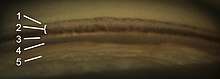 Gonioscopy of the anterior chamber angle. Labeled structures: 1. Schwalbe's line, 2. Trabecular meshwork (TM), 3. Scleral spur, 4. Ciliary body, 5. Iris
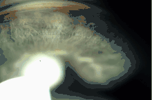
.png) Anterior chamber angle cross-section imaged by an SD-OCT.
Anterior chamber angle cross-section imaged by an SD-OCT.