Raji cell
Raji is the first continuous human cell line of hematopoietic origin.[1] The Raji cell line is widely used as a transfection host,[2]
Raji cells were derived from the B-lymphocytes of an 11-year-old Nigerian Burkitt's lymphoma male patient in 1963 by R.J.V. Pulvertaft and was further worked on by B.O. Osunkoya (University College Hospital, Ibadan, Nigeria).[3], [4]
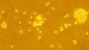
The Raji cell line is categorized as lymphoblast-like. The culture medium used to grow Raji cells is RPMI supplemented with serum. Some characteristics of Raji cells include a lack of differentiation, illustrated by the formation of large aggregations of hundreds of individual cells. The cells are relatively large in diameter (5-8 μm), have irregular indented nuclei, and almost extensive cytoplasm with free ribosomes which tend to clump.[5] Raji cells grow as single, non-motile, free-floating (non-adhesion) individuals or doublets to glass. Some cells look elongated, pear-shaped with larger, multinucleate, round cells.[5]
The Raji cell line produces an unusual strain of Epstein-Barr virus, which both transforms cord blood lymphocytes and induces early antigens in the cells. Translocations between chromosomes 8 and 22 have occurred in all three variations of the Raji cell line, but some cells synthesize immunoglobulin M with light chains of the kappa type, in contrast to the usual concordance between a translocation involving chromosome 22 and lambda chain synthesis. Both kappa genes and one lambda gene are rearranged. These findings indicate either that translocation may occur as a separate event from immunoglobulin gene rearrangement or that the proposed hierarchical sequence of immunoglobulin gene rearrangements is not always adhered to. The data also imply that in cells containing a translocation between the long arm of chromosome 8 and a chromosome bearing an immunoglobulin gene, alteration of cellular myc expression may occur regardless of the immunoglobulin gene that is expressed.[6] The cells grow in a suspension, are diploid, and are lymphoblastoid in morphology.
References
- B Fadeel (30 September 2004). "Raji revisited: cytogenetics of the original Burkitt's lymphoma cell line". Leukemia. 19 (1): 159–161. doi:10.1038/sj.leu.2403534. PMID 15457187.
- Ling Yong, CL; et al. (9 August 2014). "Microbubble-mediated sonoporation for highly efficient transfection of recalcitrant human B- cell lines". Journal of Biotechnology. 9 (8): 1081–1087. doi:10.1002/biot.201300507. PMID 24818807.
- R. J. V. Pulvertaft. A study of malignant tumours in Nigeria by short-term tissue culture. J Clin Pathol. 1965 May; 18(3): 261–273. https://pubmed.ncbi.nlm.nih.gov/14304234/
- B. O. Osunkoya. The preservation of Burkitt tumour cells at moderately low temperature. Br J Cancer. 1965 Dec; 19(4): 749–753. https://pubmed.ncbi.nlm.nih.gov/5862655/
- Cell Bank. "JCRB9012 [RAJI]". Archived from the original on May 10, 2010. Retrieved February 3, 2012.
- http://www.sciencemag.org/gca?sendit.y=9&gca=sci%3B222%2F4628%2F1094&sendit=Get%20all%20checked%20abstract%28s%29