Psittacine beak and feather disease
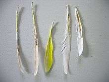
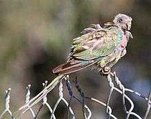
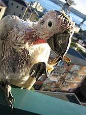
Psittacine beak and feather disease (PBFD) is a viral disease affecting all Old World and New World parrots. The causative virus–beak and feather disease virus (BFDV)—belongs to the taxonomic genus Circovirus, family Circoviridae. It attacks the feather follicles and the beak and claw matrices of the bird, causing progressive feather, claw and beak malformation and necrosis. In later stages of the disease, feather shaft constriction occurs, hampering development until eventually all feather growth stops. It occurs in an acutely fatal form and a chronic form.
| Beak and feather disease virus | |
|---|---|
| Virus classification | |
| (unranked): | Virus |
| Realm: | Monodnaviria |
| Kingdom: | Shotokuvirae |
| Phylum: | Cressdnaviricota |
| Class: | Arfiviricetes |
| Order: | Cirlivirales |
| Family: | Circoviridae |
| Genus: | Circovirus |
| Species: | Beak and feather disease virus |


Cracking and peeling of the outer layers of the claws and beak make tissues vulnerable to secondary infection. Because the virus also affects the thymus and Bursa of Fabricius, slowing lymphocyte production, immunosuppression occurs and the bird becomes more vulnerable to secondary infections. Beak fractures and necrosis of the hard palate can prevent the bird from eating.[1]
History
The ornithologist Edwin Ashby observed a flock of completely featherless red-rumped parrots (Psephotus haematonotus) in the Adelaide Hills, South Australia, in 1888. The species then disappeared from the area for several years.[2]
The condition is more prevalent in widely occurring Australian species such as the sulphur-crested cockatoo, little corella and galah.[3]
The first case of chronic PBFD was reported in a Control and Therapy article in 1972 for the University of Sydney by Ross Perry, in which he described it as "beak rot in a cockatoo".[4] Dr. Perry subsequently studied the disease and wrote extensively about its clinical features in a range of psittacine birds in a long article in which he named the disease "psittacine beak and feather disease syndrome" (PBFDS).[4] This soon became known as psittacine beak and feather disease (PBFD).[4]
The beak and feather disease virus
Beak and feather disease virus (BFDV) is a circular or icosahedral, 14–16 nm diameter, single-stranded circular DNA, non-enveloped virus with a genome size of between 1992 and 2018 nucleotides. It encodes seven open reading frames—three in the virion strand and four in the complementary strand.[5] The open reading frames have some homology to porcine circovirus (family Circoviridae), subterranean clover stunt virus and faba bean necrotic yellows virus (both family Nanoviridae).
History
It was first isolated and characterized by researchers Dr. David Pass of Murdoch University in Perth and Dr. Ross Perry from Sydney, with later work at the University of Georgia in the United States, the University of Sydney and Murdoch University in Australia, and the University of Cape Town, among other centres. The virus was originally designated PCV (psittacine circovirus), but has since been renamed beak and feather disease virus. This is due in part, to the research confirming that this virus is the cause of the disease, and in part to avoid confusion with Porcine circovirus, also called PCV.
Detection
A variety of tests for the presence of BFDV are available: standard polymerase chain reaction (PCR), quantitative PCR (qPCR) which can detect the virus in extremely small quantities, whole-genome sequencing, histology, immunohistochemical tests, and quantitative haemagglutination assays.[6]
Infection paths
PBFD is usually acquired by nestlings from their parents (vertical transmission) or from other members of the flock (horizontal transmission). The immature immune system of young birds makes them susceptible to the PBFDV. The virus may be transferred in crop secretions, in fresh or dried feces, and in feather and skin particles.
Adult birds coming into contact with the virus usually (but not always) develop resistance to it, but the virus is retained in their body and, in most cases, is excreted in feces and feather debris for the rest of their lives.
Signs of disease

The acute form of the disease is manifested by lethargy, loss of appetite, vomiting and diarrhea. Due to the severe immune system suppression, multiple secondary infections develop, causing death within two to four weeks. Typical confirmation of the acute form of the disease is by necropsy, because it progresses too quickly for the normal signs such as feather loss and beak deformity to appear.
The chronic form occurs if the bird's immune system manages to mount a reaction to the virus and any secondary infections. The characteristic feather symptoms need time to develop, as they only appear during the first moult after infection. In those species having powder down, signs may be visible immediately, as powder down feathers are continually replenished.
Threat
PBFD has the potential to become a major threat to all species of wild parrots and to modern aviculture, due to international legal and illegal bird trade. Cases of PBFD have now been reported in at least 78 psittacine species.[6] At least 38 of 50 Australian native species are affected by PBFD, both captive and in the wild. In 2004, PBFD was listed as a key threatening process by the Australian Commonwealth Government for the survival of five endangered species, including one of the few remaining species of migratory parrots, the orange-bellied parrot, of which only an estimated 3 mating pairs remained in 2017.
Treatment
There is currently no specific treatment for the virus. A vaccine is available, but only experimentally. It has not been released to the public due to the risk it poses to already exposed birds.[7][8]
Therapeutic intervention is limited to treating secondary infections. The individual bird can sometimes recover or have an acceptable quality of life if the symptoms are mild/progress slowly.
The management of the disease lies thus mostly in prevention. Every new bird that enters a pen with other birds should be quarantined first and be tested for BFDV. Birds which are known carriers should not be introduced into new pens, especially not if those contain young birds.
References
- Pyne, M. Psittacine Beak and Feather Disease. Currumbin Wildlife Sanctuary, Gold Coast. National Wildlife Rehabilitation Conference 2005.
- Ashby, E. (1921). Notes on Psephotus hematonotus, the Red-rumped Grass Parrakeet. The Avicultural Magazine Third Series, Vol. XII. pg 131.
- Borthwick, D. Threat Abatement Plan for Psittacine Beak and Feather Disease Affecting Endangered Psittacine Species. Department of the Environment and Heritage, Commonwealth of Australia. 2005.
- Perry, R.A. (197?) Proc 55, PGCVSc, University of Sydney, pp. ?-?
- Bassami MR, Berryman D, Wilcox GE, Raidal SR (1998). "Psittacine beak and feather disease virus nucleotide sequence analysis and its relationship to porcine circovirus, plant circoviruses, and chicken anaemia virus". Virology. 249 (2): 453–9. doi:10.1006/viro.1998.9324. PMID 9791035.
- Fogell, Deborah J.; Martin, Rowan O.; Groombridge, Jim J. (2016-05-05). "Beak and feather disease virus in wild and captive parrots: an analysis of geographic and taxonomic distribution and methodological trends". Archives of Virology. 161 (8): 2059–74. doi:10.1007/s00705-016-2871-2. ISSN 0304-8608. PMC 4947100. PMID 27151279.
- Beak and feather disease virus (BFDV) (micro-organism). Global Invasive Species Database. ISSG. IUCN.
- "Beak and Feather disease".
Further reading
- Pass, D. A.; Perry, R. A. (1984). "The pathology of psittacine beak and feather disease". Aust Vet J. 61 (3): 69–74. doi:10.1111/j.1751-0813.1984.tb15520.x. PMID 6743145.
- Pass, D. A.; Perry, R. A. (1985). "Psittacine beak and feather disease: An update". Aust Vet Practit. 15: 55–60.
- Raidal, S. R., et al. (2005). Development of Recombinant Proteins as a Candidate Vaccine for Psittacine Beak and Feather Disease. Final Report for the Australian Government Department of the Environment and Heritage. Murdoch University, Perth, Western Australia.