NDUFA4L2
NADH dehydrogenase (ubiquinone) 1 alpha subcomplex, 4-like 2 is a protein that in humans is encoded by the NDUFA4L2 gene.[5] The NDUFA4L2 protein is a subunit of NADH dehydrogenase (ubiquinone), which is located in the mitochondrial inner membrane and is the largest of the five complexes of the electron transport chain.[6]
| NDUFA4L2 | |||||||||||||||||||||||||
|---|---|---|---|---|---|---|---|---|---|---|---|---|---|---|---|---|---|---|---|---|---|---|---|---|---|
| Identifiers | |||||||||||||||||||||||||
| Aliases | NDUFA4L2, NUOMS, NADH dehydrogenase (ubiquinone) 1 alpha subcomplex, 4-like 2, NDUFA4, mitochondrial complex associated like 2, NDUFA4 mitochondrial complex associated like 2, COXFA4L2 | ||||||||||||||||||||||||
| External IDs | MGI: 3039567 HomoloGene: 49614 GeneCards: NDUFA4L2 | ||||||||||||||||||||||||
| |||||||||||||||||||||||||
| |||||||||||||||||||||||||
| |||||||||||||||||||||||||
| Orthologs | |||||||||||||||||||||||||
| Species | Human | Mouse | |||||||||||||||||||||||
| Entrez | |||||||||||||||||||||||||
| Ensembl | |||||||||||||||||||||||||
| UniProt | |||||||||||||||||||||||||
| RefSeq (mRNA) | |||||||||||||||||||||||||
| RefSeq (protein) | |||||||||||||||||||||||||
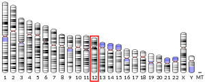
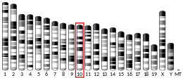
| Location (UCSC) | Chr 12: 57.23 – 57.24 Mb | Chr 10: 127.51 – 127.52 Mb | |||||||||||||||||||||||
| PubMed search | [3] | [4] | |||||||||||||||||||||||
| Wikidata | |||||||||||||||||||||||||
| |||||||||||||||||||||||||
Structure
The NDUFA4L2 gene is located on the long q arm of chromosome 12 at position 13.3 and it spans 5,860 base pairs.[5] NDUFA4L2 is a subunit of the enzyme NADH dehydrogenase (ubiquinone), the largest of the respiratory complexes. The structure is L-shaped with a long, hydrophobic transmembrane domain and a hydrophilic domain for the peripheral arm that includes all the known redox centers and the NADH binding site.[6] It has been noted that the N-terminal hydrophobic domain has the potential to be folded into an alpha helix spanning the inner mitochondrial membrane with a C-terminal hydrophilic domain interacting with globular subunits of Complex I. The highly conserved two-domain structure suggests that this feature is critical for the protein function and that the hydrophobic domain acts as an anchor for the NADH dehydrogenase (ubiquinone) complex at the inner mitochondrial membrane.[5]
Function
The human NDUFA4L2 gene codes for a subunit of Complex I of the respiratory chain, which transfers electrons from NADH to ubiquinone.[5] Initially, NADH binds to Complex I and transfers two electrons to the isoalloxazine ring of the flavin mononucleotide (FMN) prosthetic arm to form FMNH2. The electrons are transferred through a series of iron-sulfur (Fe-S) clusters in the prosthetic arm and finally to coenzyme Q10 (CoQ), which is reduced to ubiquinol (CoQH2). The flow of electrons changes the redox state of the protein, resulting in a conformational change and pK shift of the ionizable side chain, which pumps four hydrogen ions out of the mitochondrial matrix.[6]
References
- GRCh38: Ensembl release 89: ENSG00000185633 - Ensembl, May 2017
- GRCm38: Ensembl release 89: ENSMUSG00000040280 - Ensembl, May 2017
- "Human PubMed Reference:". National Center for Biotechnology Information, U.S. National Library of Medicine.
- "Mouse PubMed Reference:". National Center for Biotechnology Information, U.S. National Library of Medicine.
- "Entrez Gene: NADH dehydrogenase (ubiquinone) 1 alpha subcomplex, 4-like 2".
- Voet D, Voet JG, Pratt CW (2013). "Chapter 18". Fundamentals of biochemistry: life at the molecular level (4th ed.). Hoboken, NJ: Wiley. pp. 581–620. ISBN 978-0-470-54784-7.
This article incorporates text from the United States National Library of Medicine, which is in the public domain.


