Sponge spicule
Spicules are structural elements found in most sponges. They provide structural support and deter predators. Large spicules that are visible to the naked eye are referred to as megascleres, while smaller, microscopic ones are termed microscleres.

Structure
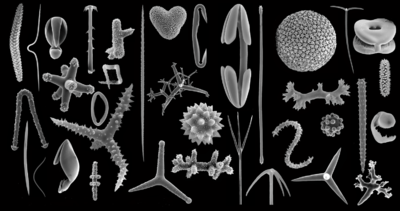
Spicules are found in a range of symmetry types.
Monaxons form simple cylinders with pointed ends. The ends of diactinal monaxons are similar, whereas monactinal monaxons have different ends: one pointed, one rounded. Diactinal monaxons are classified by the nature of their ends: oxea have pointed ends, and strongyles are rounded. Spine-covered oxea and strongyles are termed acanthoxea and acanthostrongyles, respectively.[1]:2 Monactical monaxons always have one pointed end; they are termed styles if the other end is blunt, tylostyles if their blunt end forms a knob; and acanthostyles if they are covered in spines.
Triaxons have three axes; in triods, each axis bears a similar ray; in pentacts, the triaxon has five rays, four of which lie in a single plane; and pinnules are pentacts with large spines on the non-planar ray.[1]
Tetraxons have four axes, and polyaxons more (description of types to be incorporated from [1]). Sigma-C spicules have the shape of a C.[1]
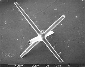
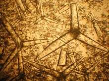
Dendroclones might be unique to extinct sponges[2] and are branching spicules that may take irregular forms, or may form structures with an I, Y or X shape.[3][4] mac/doolittle/052505/piday/3-14-2018
Spicule types

- Megascleres are large spicules measuring from 60-2000 µm and often function as the main support elements in the skeleton.
- Acanthostyles are spiny styles.
- Anatriaenes, orthotriaenes and protriaenes are triaenes[5] - megascleres with one long and three short rays.
- Strongyles are megascleres with both ends blunt or rounded.
- Styles are megascleres with one end pointed and the other end rounded.
- Tornotes are megascleres with spear shaped ends.
- Tylotes are megascleres with knobs on both ends.
- Microscleres are small spicules measuring from 10-60 µm and are scattered throughout the tissue and are not part of the main support element.
- Anisochelas are microscleres with dissimilar ends.
- Chelae are microscleres with shovel-like structures on the ends.
- Euasters are star-shaped microscleres with multiple rays radiating from a common centre.
- Forceps are microscleres bent back on themselves.
- Isochelas are microscleres with two similar ends.
- Microstrongyles are microscleres with both ends blunt or rounded.
- Oxeas are microscleres with both ends pointed.
- Oxyasters are star-shaped microscleres with thin pointed rays.
- Sigmas are "C" or "S" shaped microscleres.
- Spherasters are microscleres with multiple rays radiating from a spherical centre.[6]
Composition

Sponges can be calcareous, siliceous, or composed of spongin.
Function
The meshing of many spicules serves as the sponge’s skeleton. It provides structural support and defense against predators.
Taxonomic importance
The composition, size, and shape of spicules is one of the largest determining factors in sponge taxonomy.
Formation
Spicules are formed by sclerocytes, which are derived from archaeocytes. The sclerocyte begins with an organic filament, and adds silica to it. Spicules are generally elongated at a rate of 1-10 μm per hour. Once the spicule reaches a certain length it protrudes from the sclerocyte cell body, but remains within the cell’s membrane. On occasion, sclerocytes may begin a second spicule while the first is still in progress.[7]
Interaction with light
Research on the Euplectella aspergillum (Venus' Flower Basket) demonstrated that the spicules of certain deep-sea sponges have similar traits to Optical fibre. In addition to being able to trap and transport light, these spicules have a number of advantages over commercial fibre optic wire. They are stronger, resist stress easier, and form their own support elements. Also, the low-temperature formation of the spicules, as compared to the high temperature stretching process of commercial fibre optics, allows for the addition of impurities which improve the refractive index. In addition, these spicules have built-in lenses in the ends which gather and focus light in dark conditions. It has been theorized that this ability may function as a light source for symbiotic algae (as with Rosella racovitzae) or as an attractor for shrimp which live inside the Venus' Flower Basket. However, a conclusive decision has not been reached; it may be that the light capabilities are simply a coincidental trait from a purely structural element. [7][8][9] Spicules funnel light deep inside sea sponges.[10][11]
Applications
- Bionics application.
- Used as a kind of exfoliant in derma problem treatment.
References
- Eugenio Andri; Stefania Gerbaudo; Massimiliano Testa (2001). "13. Quaternary siliceous sponge spicules in the western Woodlark basin, southwest Pacific (ODP LEG 180)" (PDF). In Huchon, P.; Taylor, B.; Klaus, A (eds.). Proceedings of the Ocean Drilling Program, Scientific Results. 180. pp. 1–8.
- Komal Sutar and Bhavika Bhoir. "Study of the sponge spicules from the coastline of Raigad district, Maharashtra, India" (PDF). vpmthane.org/.
- Rigby, J. K.; Boyd, D. W. (2004). "Sponges from the Park City Formation (Permian) of Wyoming". Journal of Paleontology. 78: 71–76. doi:10.1666/0022-3360(2004)078<0071:SFTPCF>2.0.CO;2. ISSN 0022-3360.
- Bingli, L.; Rigby, J.; Zhongde, Z. (2003). "Middle Ordovician Lithistid Sponges from the Bachu-Kalpin Area, Xinjiang, Northwestern China". Journal of Paleontology. Paleontological Society. 77 (3): 430–441. doi:10.1666/0022-3360(2003)077<0430:MOLSFT>2.0.CO;2. JSTOR 4094792.
- Boury-Esnault, Nicole, and Klaus Rutzler, editors. Thesaurus of Sponge Morphology. Smithsonian Contributions to Zoology, number 596, 55 pages, 305 figures, 1997. https://www.portol.org/thesaurus
- Glossary: Rosario Beach Marine Laboratory
- Imsiecke G, Steffen R, Custodio M, Borojevic R, Müller WE (August 1995). "Formation of spicules by sclerocytes from the freshwater sponge Ephydatia muelleri in short-term cultures in vitro". In Vitro Cell Dev Biol Anim. 31 (7): 528–35. doi:10.1007/BF02634030. PMID 8528501.
- Aizenburg, Joanna; et al. (2004). "Biological glass fibers: Correlation between optical and structural properties". Proceedings of the National Academy of Sciences. 101 (10): 3358–3363. Bibcode:2004PNAS..101.3358A. doi:10.1073/pnas.0307843101. PMC 373466. PMID 14993612.
- Aizenberg, J.; Sundar, V. C.; Yablon, A. D.; Weaver, J. C.; Chen, G. (March 2004). "Biological glass fibers: Correlation between optical and structural properties". Proceedings of the National Academy of Sciences. 101 (10): 3358–3363. Bibcode:2004PNAS..101.3358A. doi:10.1073/pnas.0307843101. PMC 373466. PMID 14993612.
- Brümmer, Franz; Pfannkuchen, Martin; Baltz, Alexander; Hauser, Thomas; Thiel, Vera (2008). "Light inside sponges". Journal of Experimental Marine Biology and Ecology. 367 (2): 61–64. doi:10.1016/j.jembe.2008.06.036.
- "Nature's 'fibre optics' experts". BBC. 2008-11-10. Retrieved 2008-11-10.