Immune electron microscopy
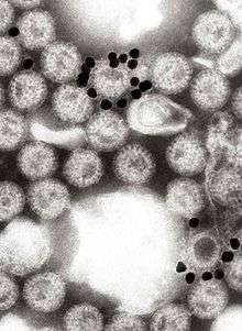
Immune electron microscopy is a variation of electron microscopy. It is applied for diagnosis of many viral infections.[1] A difficult procedure,[2] IEM was developed as a diagnostic aid for detecting and identifying transmissible gastroenteritis virus and rotavirus (reovirus-like agent) in fecal and intestinal contents from cases of gastroenteritis in young pigs.[3] IEM is one of the fastest and most sensitive methods for the detection and diagnosis of viruses. This technique is based on formation of immune complexes of the virus with its corresponding antibody.[4]
Procedure

IEM is used to localize molecules at the ultrastructural level by labeling them with specific antibodies, which are visualized by electron-opaque markers (colloidal gold particles) attached to them. The effect is to produce an electron-dense label at the site of the antigen-antibody reaction. When the antigen is located inside of the cell, then transmission electron microscopy is required to see it. The labeling can be done pre-embedding or post-embedding. When the antigen in question is located on the surface of the specimen, scanning electron microscopy can be used to see it. The electron dense label can then be viewed using the back scatter image on an appropriately equipped microscope.[5]
Application
Although IEM is a specific, sensitive, and quantitative assay for hepatitis A antigen and antibody to HA Ag (anti-HA), its practicality for large scale testing is limited. In 1973, Feinstone et al. used IEM to examine stool specimens taken from prison volunteers infected with the Willowbrook MS-1 strain of hepatitis A virus.[6] A well-documented and a highly sensitive virus detection technique, IEM has also been found to be highly effective for potato viruses.[7] IEM also has been used for detecting plant viruses.[8]
References
- Orthopoxviruses Pathogenic for Humans. 2006-06-09. ISBN 9780387253060.
- Bradley, David E. (1984). "Characteristics and Function of Thick and Thin Conjugative Pili Determined by Transfer-derepressed Plasmids of Incompatibility Groups It, Ia, Is, B, K and Z" (PDF). Microbiology. 130 (6): 1489–1502. doi:10.1099/00221287-130-6-1489. PMID 6148378. Retrieved 1 October 2018.
- Saif, LJ; Bohl, EH; Kohler, EM; Hughes, JH (1977). "Immune electron microscopy of transmissible gastroenteritis virus and rotavirus (reovirus-like agent) of swine". American Journal of Veterinary Research. 38 (1): 13–20. PMID 189646.
- Katz, D; Straussman, Y; Shahar, A; Kohn, A (1980). "Solid-phase immune electron microscopy (SPIEM) for rapid viral diagnosis". Journal of Immunological Methods. 38 (1–2): 171–4. doi:10.1016/0022-1759(80)90341-5. PMID 7452001.
- "Immuno-Electron Microscopy". umassmed.edu. 2013-11-02. Retrieved 1 October 2018.
- Dienstag, Jules L.; Purcell, Robert H. (1977). "Recent advances in the identification of hepatitis viruses*" (PDF). Postgraduate Medical Journal. 53 (621): 364–373. doi:10.1136/pgmj.53.621.364. Retrieved 1 October 2018.
- Gars, I. D.; Paul Khurana, S. M. (1991). Horticulture — New Technologies and Applications. Current Plant Science and Biotechnology in Agriculture. 12. pp. 329–336. doi:10.1007/978-94-011-3176-6_52. ISBN 978-94-010-5401-0.
- Shukla, D. D.; Gough, K. H. (1979). "The Use of Protein A, from Staphylococcus aureus, in Immune Electron Microscopy for Detecting Plant Virus Particles" (PDF). Journal of General Virology. 45 (2): 533–536. doi:10.1099/0022-1317-45-2-533. Retrieved 1 October 2018.