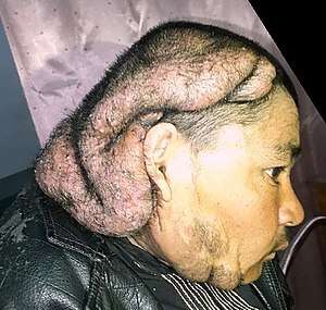
Cutis verticis gyrata
Cutis verticis gyrata is a medical condition usually associated with thickening of the scalp.[1] People show visible folds, ridges or creases on the surface of the top of the scalp.[2] The number of folds can vary from two to roughly ten and are typically soft and spongy. These folds cannot be corrected with pressure. The condition typically affects the central and rear regions of the scalp, but sometimes can involve the entire scalp.
| Cutis verticis gyrata | |
|---|---|
| Other names | CVG |
 | |
Hair loss can occur over time where the scalp thickens, though hair within any furrows remains normal. Thus far, due to the (apparent) rarity of the condition, limited research exists and causes are as yet undetermined. What is known, is that the condition is not exclusively congenital.
The condition was first reported by Jean-Louis-Marc Alibert in 1837,[3] who called it cutis sulcata.[4] A clinical description of the condition was provided by Robert in 1843[5] and it was named by Paul Gerson Unna in 1907.[6] It has also been called Robert-Unna syndrome, bulldog scalp, corrugated skin, cutis verticis plicata, and pachydermia verticis gyrata.[7]
Cause
At this time, causes are unknown, but it is believed to not be congenital.
Diagnosis
There is no clinical diagnosis for CVG as cases are rarely seen and are often comorbid with other conditions.
Classifications
CVG is classified according to the presence, or lack of underlying cause. Studies suggest that CVG often occurs in individuals in a secondary form to other ailments. However, the condition can also be present on its own. CVG can be classified into two forms: ‘primary’ (essential and non-essential) and ‘secondary’.[8]
The classifications are:
- Primary essential
- Primary non-essential
- Secondary
Primary essential CVG is where the cause of the condition in unknown. It has no other associated abnormalities. This occurs mainly in men, with a male:female ratio of 5:1 or 6:1, and develops during or soon after puberty. Because of the slow progression of the condition, which usually occurs without symptom, it often passes unnoticed in the early stage.[9]
Primary non-essential CVG can be associated with neuropsychiatric disorders including cerebral palsy, epilepsy, seizures, and ophthalmologic abnormalities, most commonly cataracts.
Secondary CVG occurs as a consequence of a number of diseases or drugs that produce changes in scalp structure. These include: acromegaly (excessive growth hormone levels due to pituitary gland tumours), and theoretically, the use of growth hormone itself or the use of drugs that mimic the effect of growth hormone (such as GHRP-6 and CJC-1295). It may also arise in association with melanocytic naevi (moles), birthmarks (including connective tissue naevi, fibromas and naevus lipomatosus), and inflammatory processes (e.g. eczema, psoriasis, Darier disease, folliculitis, impetigo, atopic dermatitis, acne).
Treatment
The only current medical treatment for this condition is limited to plastic surgery with excision of the folds by means of scalp reduction/surgical resection. Scalp subcision has also been suggested as a treatment.[10] Additional suggestions also include injections of a dermal filler (e.g. (poly-L-lactic acid)).[11]
See also
- Skin lesion
- List of cutaneous conditions
References
Notes
- James, William; Berger, Timothy; Elston, Dirk (2005). Andrews' Diseases of the Skin: Clinical Dermatology (10th ed.). Saunders. p. 572. ISBN 978-0-7216-2921-6.
- Rapini, Ronald P.; Bolognia, Jean L.; Jorizzo, Joseph L. (2007). Dermatology: 2-Volume Set. St. Louis: Mosby. p. 1502. ISBN 978-1-4160-2999-1.
- Tan O, Ergen D (July 2006). "Primary essential cutis verticis gyrata in an adult female patient: a case report". J. Dermatol. 33 (7): 492–5. doi:10.1111/j.1346-8138.2006.00116.x. PMID 16848824.
- http://scholar.valpo.edu/cgi/viewcontent.cgi?article=1040&context=jmms -- See page 82 of J Mind Med Sci. 2016; 3(1): 80-87.
- "Cutis Verticis Gyrata: Background, Pathophysiology, Etiology". February 2019. Cite journal requires
|journal=(help) - Unna PG. Cutis verticis gyrata. Monatschr Prakt Derm. 1907. 45:227-33.
- Levine, Norman (2004). Dermatology Therapy A-Z Essentials. Berlin New York: Springer. p. 166. ISBN 978-3-540-00864-4.
- Radwanski, Henrique N.; Rocha Almeida, Marcelo Wilson; Pitanguy, Ivo (2009). "Primary essential cutis verticis gyrata – a case report". Journal of Plastic, Reconstructive & Aesthetic Surgery. 62 (11): e430–e433. doi:10.1016/j.bjps.2008.06.062. ISSN 1748-6815. PMID 18951076.
- Okamoto K, Ito J, Tokiguchi S, Ishikawa K, Furusawa T, Sakai K (October 2001). "MRI in essential primary cutis verticis gyrata". Neuroradiology. 43 (10): 841–4. doi:10.1007/s002340100591. PMID 11688700. S2CID 293444.
- Hsu YJ, Chang YJ, Su LH, Hsu YL (March 2009). "Using novel subcision technique for the treatment of primary essential cutis verticis gyrata". Int. J. Dermatol. 48 (3): 307–9. doi:10.1111/j.1365-4632.2009.03927.x. PMID 19261024.
- Unusual and rare complication described in San Francisco | CATIE - Canada's source for HIV and hepatitis C information
Bibliography
- Nguyen NQ (October 2003). "Cutis verticis gyrata". Dermatol. Online J. 9 (4): 32. PMID 14594605.