Cotesia congregata
Cotesia congregata is a parasitoid wasp of the genus Cotesia. The genus is particularly noted for its use of polydnaviruses. Parasitoids are distinct from true parasites in that a parasitoid will ultimately kill its host or otherwise sterilize it.
| Cotesia congregata | |
|---|---|
 | |
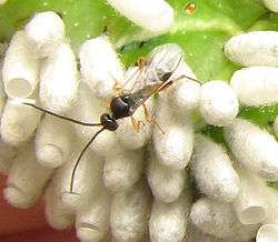
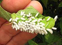
| C. congregata on hornworm Manduca sexta | |
| Male C. congregata courtship song | |
| Scientific classification | |
| Kingdom: | Animalia |
| Phylum: | Arthropoda |
| Class: | Insecta |
| Order: | Hymenoptera |
| Family: | Braconidae |
| Genus: | Cotesia |
| Species: | C. congregata |
| Binomial name | |
| Cotesia congregata (Say, 1836) | |
| Synonyms | |

Life cycle
Adult wasps lay their eggs in tobacco hornworm (Manduca sexta) larvae in their 2nd or 3rd instar (each instar is a stage between moltings, i.e. the second instar is the life stage after the first molt and before the second molting) and at the same time injects symbiotic viruses into the hemocoel of the host along with some venom. The viruses knock down the internal defensive responses of the hornworm. The eggs hatch in the host hemocoel within two to three days and simultaneously release special cells from the egg's serosa. These special cells, called teratocytes, grow to become giant cells visible to the naked eye. The teratocytes secrete hormones which work in tandem with the virus and the wasp venom to arrest the development of the host.[2] Following hatching in the caterpillar, the wasp larvae will undergo 2 molts inside the host caterpillar’s hemocoel and, after 12 to 16 days post oviposition, the 3rd instar wasp larvae will emerge from the caterpillar and spin cocoons from which the adult wasps fly about 4 to 8 days later.[3]
This insect has the shortest flagellated spermatozoa in animals, being 6.6 µm long (nucleus and flagellum), 8800 times shorter than the longest ones (Drosophila bifurca).[4]
Wasp pupae may themselves be parasitized by chalcid wasps of the genus Hypopteromalus.[5]
Polydnavirus symbiosis
An important aspect of the symbiotic polydnavirus is the fact that the virus does not and cannot replicate on its own- it does not contain the genes necessary to replicate itself. Instead, the genes that code for the virus are contained within the genome of the wasp. The wasp contains special cells called calyx cells within its ovary, which in females will produce the virion particles. Male wasps contain the viral sequence, but do not have the capacity to produce it. The proteins and genetic payload of the virus are produced by these cells, and the virions are assembled within the nucleus of these cells. As the female matures, the nuclear membrane will dissolve, followed by the cell membrane, releasing the virions and cell debris into the lumen of the oviduct. Phagocytic cells will clean up the debris, and the virions will be injected into the host along with eggs and venom upon oviposition.[6][7]
An average female wasp will produce over 600 ng of viral DNA in each ovary, more than enough for her lifetime. An average female will lay 1757 +/- 945 eggs in her lifetime, and only 0.1 ng of viral DNA is injected per egg.[8][9]
Effects of the virus on the host
The polydnavirus will severely interfere with development of the host, Manduca sexta. Infected hosts will not undergo metamorphosis and will instead reach extremely high weight and sometimes reach a supernumerary sixth instar. Upon reaching the fifth instar, the caterpillar will enter a wandering stage, as is typical, but will not progress further and will not form a cocoon. The onset of the wandering stage is temporally delayed, as well.[10]
Certain neuropeptides were found to accumulate in the neurosecretory system of the host, which were correlated to a change in molting behavior. Similar accumulation has been found in neural system of starving, unparasitized caterpillars, but not nearly to the same extent. The polydnavirus was found to inhibit development of the optic lobe of the host, causing morphological differences. One known hormone which has been focused upon, prothoracictropic hormone (PTTH), was of particular interest. It is accumulated far more heavily in parasitized and starved hosts than in normal larvae. Other proteins found to increase in neurosecretory cells in both starved and parasitized larvae are: bombyxin, allatotropin, diuretic hormone, FMRFamide, and proctolin. Other proteins were found in increased concentration in hosts from which the wasps had already emerged, such as eclosion hormone and adipokinetic hormone.[11]
The polydnavirus disallows these proteins from being released into the nervous system, instead causing them to accumulate in neurosecretory cells. Specifically with PTTH, due to the accumulation, is not released in sufficient amounts to stimulate the synthesis of ecdysteroids by the prothoracic glands, which will prevent subsequent development of the larvae. These hormones also allow the parasitized larva to survive longer without food or water, due to a slowdown of diuresis (urine production) and gut purge. This would help the larva to conserve water. Starved larvae can also ultimately molt and pupate if they are large enough, but this can be explained by the temporal difference in the beginning of accumulation. The mechanism behind neuropeptide accumulation is unknown. The polydnavirus is not the only factor affecting development of the host; teratocytes will have a similar effect, and it is likely that a large combination of different factors is needed to replicate the biological effects of parasitization.[12]
Another extremely important effect of the virus is the suppression of the immune system of the host. This is accomplished by altering the behavior of host hemocytes, including inducing apoptosis. Within 24 hours of oviposition, the host is unable to encapsulate any antigen which enters its body, preventing it from attacking the wasp larvae. The host immune system returns to normal after 8 days, by which time the wasp larvae have already built up an immunity to the immune system. Larvae which have reached 8 days old are able to survive and eclose when transplanted into a new host which has not been exposed to the virus. However, these transplanted larvae will exhibit a mortality rate of 50%.[13]
The wasp also injects venom along with the eggs and virus particles. The venom on its own will have a negligible effect on the host, but will enhance the effects of the virus when both are present.[14]
Genetic description of the virus
The Cotesia congregata bracovirus has one of the largest genomes known of any virus (567,670 base pairs), and is largely composed of introns, which is rare for a virus; 70% of the DNA is noncoding. The genome is arranged in 30 circles of DNA, which range in size from 5,000 to 40,000 base pairs. Of the 30, 29 circles code for at least one protein product. The genome is composed of 66% A-T residues.[15] The top gene products are:
- PTP proteins (protein tyrosine phosphatases), which will dephosphorylate tyrosine AA's on regulatory proteins. The PTP will interfere with certain cytoskeleton dynamics, which would be helpful in avoiding encapsulation. The PTPs found are more closely related to cellular PTPs than those found in viruses.
- The second group of proteins are ank proteins, those with ankyrin repeat motifs. These are known to inhibit immune responses in vertebrates.
- The third group of proteins are cysteine-rich proteins, are extremely similar to proteins excreted by the wasp teratocytes. These are suspected to inhibit translation of storage proteins such as arylphorin, which would leave more resources free for the parasite larvae.
- The fourth group are cystatin proteins, which will inhibit cysteine proteases. These will inhibit the breakdown of the proteins in group 3. Cystatins are not a gene product previously found in viruses to date. They also likely have an immuno-suppressive function, based on similar proteins which have been found in parasitic nematodes.
The rest of the protein products have no known homologs and their function is not known. Much of what has been discovered makes it difficult to place the virus in a phylogenetic niche, and lends support to the theory that the virus was assembled, rather than evolved. The most closely related viruses are the nudiviruses and their baculovirus relatives, although this relatedness goes back approximately 75 million years.[16]
References
- Say, T., 1836. Description of new species of North American Hymenoptera, and observations on some already described. Boston Journal of Natural History, Vol. 1, p. 262 (https://www.biodiversitylibrary.org/item/100882#page/265/mode/1up)
- Beckage, Nancy E.; Tan, Frances F.; Schleifer, Kathleen W.; Lane, Roni D.; Cherubin, Lisa L. (1 January 1994). "Characterization and biological effects ofcotesia congregata polydnavirus on host larvae of the tobacco hornworm, manduca sexta". Archives of Insect Biochemistry and Physiology. 26 (2–3): 165–195. doi:10.1002/arch.940260209.
- de Buron, Isaure; Beckage, Nancy E. (May 1992). "Characterization of a polydnavirus (PDV) and virus-like filamentous particle (VLFP) in the braconid wasp Cotesia congregata (Hymenoptera: Braconidae)". Journal of Invertebrate Pathology. 59 (3): 315–327. doi:10.1016/0022-2011(92)90139-U.
- Uzbekov, Rustem; Burlaud-Gaillard, Julien; Garanina, Anastasiia S.; Bressac, Christophe (2017). "The length of a short sperm: Elongation and shortening during spermiogenesis in Cotesia congregata (Hymenoptera, Braconidae)". Arthropod Structure & Development. 46 (2): 265–273. doi:10.1016/j.asd.2016.11.011.
- Orr, David (2018). "Hyperparasitoids". North Carolina Cooperative Extension. Retrieved 5 August 2020.
- Marti, D. (January 2003). "Ovary development and polydnavirus morphogenesis in the parasitic wasp Chelonus inanitus. I. Ovary morphogenesis, amplification of viral DNA and ecdysteroid titres". Journal of General Virology. 84 (5): 1141–1150. doi:10.1099/vir.0.18832-0.
- Wyler, T. (January 2003). "Ovary development and polydnavirus morphogenesis in the parasitic wasp Chelonus inanitus. II. Ultrastructural analysis of calyx cell development, virion formation and release". Journal of General Virology. 84 (5): 1151–1163. doi:10.1099/vir.0.18830-0.
- Lavine, M.D.; Beckage, N.E. (January 1996). "Temporal pattern of parasitism-induced immunosuppression in Manduca sexta larvae parasitized by Cotesia congregata". Journal of Insect Physiology. 42 (1): 41–51. doi:10.1016/0022-1910(95)00081-X.
- Zitnan, D; Kingan, TG; Kramer, SJ; Beckage, NE (May 22, 1995). "Accumulation of neuropeptides in the cerebral neurosecretory system of Manduca sexta larvae parasitized by the braconid wasp Cotesia congregata". The Journal of Comparative Neurology. 356 (1): 83–100. doi:10.1002/cne.903560106. PMID 7629311.
- Zitnan, D; Kingan, TG; Kramer, SJ; Beckage, NE (May 22, 1995). "Accumulation of neuropeptides in the cerebral neurosecretory system of Manduca sexta larvae parasitized by the braconid wasp Cotesia congregata". The Journal of Comparative Neurology. 356 (1): 83–100. doi:10.1002/cne.903560106. PMID 7629311.
- Lavine, M.D.; Beckage, N.E. (January 1996). "Temporal pattern of parasitism-induced immunosuppression in Manduca sexta larvae parasitized by Cotesia congregata". Journal of Insect Physiology. 42 (1): 41–51. doi:10.1016/0022-1910(95)00081-X.
- Beckage, Nancy E.; Tan, Frances F.; Schleifer, Kathleen W.; Lane, Roni D.; Cherubin, Lisa L. (1 January 1994). "Characterization and biological effects ofcotesia congregata polydnavirus on host larvae of the tobacco hornworm, manduca sexta". Archives of Insect Biochemistry and Physiology. 26 (2–3): 165–195. doi:10.1002/arch.940260209.
- Espagne, E. (8 October 2004). "Genome Sequence of a Polydnavirus: Insights into Symbiotic Virus Evolution". Science. 306 (5694): 286–289. Bibcode:2004Sci...306..286E. doi:10.1126/science.1103066. PMID 15472078.
- Espagne, E. (8 October 2004). "Genome Sequence of a Polydnavirus: Insights into Symbiotic Virus Evolution". Science. 306 (5694): 286–289. Bibcode:2004Sci...306..286E. doi:10.1126/science.1103066. PMID 15472078.