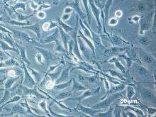
Chinese hamster ovary cell
Chinese hamster ovary (CHO) cells are an epithelial cell line derived from the ovary of the Chinese hamster, often used in biological and medical research and commercially in the production of therapeutic proteins.[1] They have found wide use in studies of genetics, toxicity screening, nutrition and gene expression, particularly to express recombinant proteins. CHO cells are the most commonly used mammalian hosts for industrial production of recombinant protein therapeutics.[1]

History
Chinese hamsters had been used in research since 1919, where they were used in place of mice for typing pneumococci. They were subsequently found to be excellent vectors for transmission of kala-azar (visceral leishmaniasis), facilitating Leishmania research.
In 1948, the Chinese hamster was first used in the United States for breeding in research laboratories. In 1957, Theodore T. Puck obtained a female Chinese hamster from Dr. George Yerganian's laboratory at the Boston Cancer Research Foundation and used it to derive the original Chinese hamster ovary (CHO) cell line. Since then, CHO cells have been a cell line of choice because of their rapid growth in suspension culture and high protein production.[2]
Having a very low chromosome number (2n=22) for a mammal, the Chinese hamster is also a good model for radiation cytogenetics and tissue culture.[3]
Properties
All CHO cell lines are deficient in proline synthesis.[4] Also, CHO cells do not express the epidermal growth factor receptor (EGFR), which makes them ideal in the investigation of various EGFR mutations.[5]
Variants
Since the original CHO cell line was described in 1956, many variants of the cell line have been developed for various purposes.[4] In 1957, CHO-K1 was generated from a single clone of CHO cells,[6] CHO-K1 was mutagenized with ethyl methanesulfonate to generate a cell line lacking dihydrofolate reductase (DHFR) activity, referred to as CHO-DXB11 (also referred to as CHO-DUKX).[7] However, these cells, when mutagenized, could revert to DHFR activity, making their utility for research somewhat limited.[7] Subsequently, CHO cells were mutagenized with gamma radiation to yield a cell line in which both alleles of the DHFR locus were completely eliminated, termed CHO-DG44[8] These DHFR-deficient strains require glycine, hypoxanthine, and thymidine for growth.[8] Cell lines with mutated DHFR are useful for genetic manipulation as cells transfected with a gene of interest along with a functional copy of the DHFR gene can easily be screened for in thymidine-lacking media. Due to this, CHO cells lacking DHFR are the most widely used CHO cells for industrial protein production.[4]
Genetic manipulation
Much of the genetic manipulation done in CHO cells is done in cells lacking DHFR enzyme. This genetic selection scheme remains one of the standard methods to establish transfected CHO cell lines for the production of recombinant therapeutic proteins. The process begins with the molecular cloning of the gene of interest and the DHFR gene into a single mammalian expression system. The plasmid DNA carrying the two genes is then transfected into cells, and the cells are grown under selective conditions in a thymidine-lacking medium. Surviving cells will have the exogenous DHFR gene along with the gene of interest integrated in its genome.[9][10] The growth rate and the level of recombinant protein production of each cell line varies widely. To obtain a few stably transfected cell lines with the desired phenotypic characteristics, evaluating several hundred candidate cell lines may be necessary.
The CHO and CHO-K1 cell lines can be obtained from a number of biological resource centres such as the European Collection of Cell Cultures, which is part of the Health Protection Agency Culture Collections. These organizations also maintain data, such as growth curves, timelapse videos of growth, images, and subculture routine information.[11]
Industrial use
CHO cells are the most common mammalian cell line used for mass production of therapeutic proteins.[1] They can produce recombinant protein on the scale of 3-10 grams per liter of culture.[4] CHO cells are also suitable for human applications, as they allow post-translational modifications to recombinant proteins which can function in humans.[12]
See also
| Wikimedia Commons has media related to CHO cells. |
References
- Wurm FM (2004). "Production of recombinant protein therapeutics in cultivated mammalian cells". Nature Biotechnology. 22 (11): 1393–1398. doi:10.1038/nbt1026. PMID 15529164.
- Fanelli, Alex (2016). "CHO Cells". Retrieved 28 November 2017.
- Tjio J. H.; Puck T. T. (1958). "Genetics of somatic mammalian cells. II. chromosomal constitution of cells in tissue culture". J. Exp. Med. 108 (2): 259–271. doi:10.1084/jem.108.2.259. PMC 2136870. PMID 13563760.
- Wurm FM; Hacker D (2011). "First CHO genome". Nature Biotechnology. 29 (8): 718–20. doi:10.1038/nbt.1943. PMID 21822249.
- Ahsan, A.; S. M. Hiniker; M. A. Davis; T. S. Lawrence; M. K. Nyati (2009). "Role of Cell Cycle in Epidermal Growth Factor Receptor Inhibitor-Mediated Radiosensitization". Cancer Research. 69 (12): 5108–5114. doi:10.1158/0008-5472.CAN-09-0466. PMC 2697971. PMID 19509222.
- Lewis NE; Liu X; Li Y; Nagarajan H; Yerganian G; O'Brien E; et al. (2013). "Genomic landscapes of Chinese hamster ovary cell lines as revealed by the Cricetulus griseus draft genome". Nature Biotechnology. 31 (8): 759–765. doi:10.1038/nbt.2624. PMID 23873082.
- Urlaub G; Chasin LA (July 1980). "Isolation of Chinese hamster cell mutants deficient in dihydrofolate reductase activity". Proceedings of the National Academy of Sciences of the United States of America. 77 (7): 4216–4220. doi:10.1073/pnas.77.7.4216. PMC 349802. PMID 6933469.
- Urlaub G; Kas E; Carothers AD; Chasin LA (June 1983). "Deletion of the diploid dihydrofolate reductase locus from cultured mammalian cells". Cell. 33 (2): 405–412. doi:10.1016/0092-8674(83)90422-1. PMID 6305508.
- Lee F; Mulligan R; Berg P; Ringold G (19 November 1981). "Glucocorticoids regulate expression of dihydrofolate reductase cDNA in mouse mammary tumour virus chimaeric plasmids". Nature. 294 (5838): 228–232. doi:10.1038/294228a0. PMID 6272123.
- Kaufman RJ; Sharp PA (25 August 1982). "Amplification and expression of sequences cotransfected with a modular dihydrofolate reductase complementary DNA gene". Journal of Molecular Biology. 159 (4): 601–621. doi:10.1016/0022-2836(82)90103-6. PMID 6292436.
- "General Cell Collection: CHO-K1". Hpacultures.org.uk. 2000-01-01. Retrieved 2013-05-21.
- Tingfeng, Lai; et al. (2013). "Advances in Mammalian Cell Line Development Technologies for Recombinant Protein Production". Pharmaceuticals. 6 (5): 579–603. doi:10.3390/ph6050579. PMC 3817724. PMID 24276168.
External links
- Chinese Hamster Genome Database
- https://web.archive.org/web/20131213154144/http://pef.aibn.uq.edu.au/wordpress/wp-content/blogs.dir/1/files/Support/Mammalian/Literature/Recombinant_Protein_Therapeutics_from_CHO_Cells-20_years_and_counting_Jayapal.pdf
- Puck TT, Cieciura SJ, Robinson A (December 1958). "Genetics of somatic mammalian cells. III. Long-term cultivation of euploid cells from human and animal subjects". J. Exp. Med. 108 (6): 945–56. doi:10.1084/jem.108.6.945. PMC 2136918. PMID 13598821.
- Cellosaurus entry for CHO
- Cellosaurus entry for CHO-K1
- Cellosaurus entry for CHO-DG44
- Cellosaurus entry for CHO-DXB11