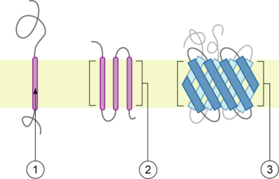
Bitopic protein
Bitopic proteins (also known as single-pass or single-spanning proteins) are transmembrane proteins that span the lipid bilayer only one time.[1] These proteins may constitute up to 50% of all transmembrane proteins, depending on the organism, and contribute significantly to the network of interactions between different proteins in cells, including interactions via transmembrane helices.[2] They usually include one or several water-soluble domains situated at the different sides of biological membranes, for example in single-pass transmembrane receptors. Some of them are small and serve as regulatory or structure-stabilizing subunits in large multi-protein transmembrane complexes, such as photosystems or the respiratory chain.

The membrane is represented in light-brown.
Topology-based classification
Bitopic proteins are classified into 4 types, depending on their transmembrane topology and location of the transmembrane helix in the amino acid sequence of the protein. According to Uniprot[3]:
- Type I is bitopic protein with its N-terminus on the extracellular side of the membrane and removed signal peptide;
- Type II is bitopic protein with its N-terminus on the cytoplasmic side of the membrane and the transmembrane helix located close to the N-terminus, where it works as an anchor;
- Type III is bitopic protein with its N-terminus on the extracellular side of the membrane and no signal peptide;
- Type IV is bitopic protein with its N-terminus on the cytoplasmic side of the membrane, and the transmembrane helix located close to the C-terminus, where it works as an anchor.
Hence type I proteins are anchored to the lipid membrane with a stop-transfer anchor sequence and have their N-terminal domains targeted to the ER lumen during synthesis (and the extracellular space, if mature forms are located on plasmalemma). Type II and III are anchored with a signal-anchor sequence, with type II being targeted to the ER lumen with its C-terminal domain, while type III have their N-terminal domains targeted to the ER lumen. Type IV is subdivided into IV-A, with their N-terminal domains targeted to the cytosol and IV-B, with an N-terminal domain targeted to the lumen.[4] The implications for the division in the four types are especially manifest at the time of translocation and ER-bound translation, when the protein has to be passed through the ER membrane in a direction dependent on the type.
Databases
- Membranome database is a database of bitopic proteins from several model organisms.
- Bitopic proteins in OPM database
References
- Membrane Structural Biology: With Biochemical and Biophysical Foundations, by Mary Luckey, 2014, Cambridge University Press, page 91.
- Zviling, Moti; Kochva, Uzi; Arkin, Isaiah T. (2007). "How important are transmembrane helices of bitopic membrane proteins?". Biochimica et Biophysica Acta (BBA) - Biomembranes. 1768 (3): 387–392. doi:10.1016/j.bbamem.2006.11.019. PMID 17258687.
- Topology definitions in Uniprot, see ,
- Harvey Lodish etc.; Molecular Cell Biology, Sixth edition, p.546