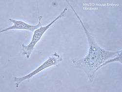
3T3 cells
3T3 cells come from a cell line established in 1962 by two scientists then at the Department of Pathology in the New York University School of Medicine, George Todaro and Howard Green. Todaro and Green originally obtained their 3T3 cells from Swiss albino mouse embryo tissue.[1] After landing a principal investigator position at the National Cancer Institute in Bethesda, Maryland Tadaro repeated the isolation procedure from the NIH Swiss mouse embryo with his students and established NIH/3T3 cell line. [2]
Nomenclature
The '3T3' designation refers to the abbreviation of "3-day transfer, inoculum 3×10⁵ cells." This cell line was originally established from the primary mouse embryonic fibroblast cells that were cultured by the designated protocol, so-called '3T3 protocol'. The primary mouse embryonic fibroblast cells were transferred (the "T") every 3 days (the first "3"), and inoculated at the rigid density of 3×10⁵ cells per 20 cm2 dish (the second "3") continuously [2]. The spontaneously immortalized cells with stable growth rate were established after 20 to 30 generations in culture, and then named '3T3' cells. Specifically, "3T3-L1" is one of the current lines.
Characteristics
Swiss 3T3 can be inhibited by temazepam and other benzodiazepines. These cells are also contact inhibited. The cells are sensitive to sarcoma virus and leukemia virus focus formation. 3T3 cells can be transformed with SV40 and some other polyomaviruses.[3]
Culture characteristics
Adherent cells grow as a monolayer. A confluent monolayer yields 40000 cells/cm2. [4]
Expression
Lysophosphatidylcholine (lyso-PC) induces AP-1 activity and c-jun N-terminal kinase activity (JNK1) by a protein kinase C-independent pathway. [5]
Cytogenetics
3T3 mouse cells are hypertriploid. The modal chromosome number is 68, which occurs in 30% of cells. Higher ploidies occur at a much lower rate of 2.4%.[4]
Uses
3T3 cells are often used in the cultivation of keratinocytes, with the 3T3 cells secreting growth factors favourable to these kinds of cells.
References
- Todaro, GJ; Green, H (1963). "Quantitative studies of the growth of mouse embryo cells in culture and their development into established lines". J. Cell Biol. 17 (2): 299–313. doi:10.1083/jcb.17.2.299. PMC 2106200. PMID 13985244.
- "Generating the 3T3 cell line, the oncogene hypothesis and horses". www.asbmb.org. Retrieved 2020-07-28.
- NIH 3T3 Cell Line Origins, Characteristics, Expression, and Cytogenetics
- "3T3-Swiss albino ATCC ® CCL-92™ Mus musculus embryo". www.atcc.org. Retrieved 2020-07-28.
- Fang, X.; Gibson, S.; Flowers, M.; Furui, T.; Bast, R. C.; Mills, G. B. (1997-05-23). "Lysophosphatidylcholine stimulates activator protein 1 and the c-Jun N-terminal kinase activity". The Journal of Biological Chemistry. 272 (21): 13683–13689. doi:10.1074/jbc.272.21.13683. ISSN 0021-9258. PMID 9153219.
External links
| Wikimedia Commons has media related to 3T3 cells. |
- Cellosaurus entry for 3T3
- Nikon MicroscopyU Digital Video Gallery: 3T3 Cell Motility - a set of films of 3T3 cells in culture