
Background and Identification
Fluoroscopy is an imaging technique that uses X-rays to produce real-time moving images of the interior of an object or body. Fluoroscopes are primarily used in medical imaging and allow physicians to see the internal structure of a patient. Fluoroscopes allow doctors to see things like the pumping action of the heart or the motion of swallowing. This is useful for diagnosis and therapy and is used in general radiology, interventional radiology, and image-guided surgery.
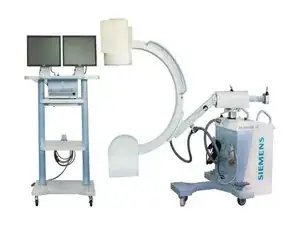
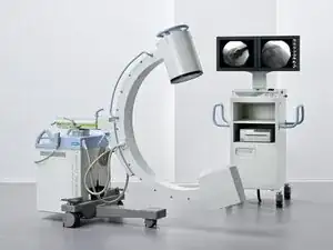
Fluoroscopes consist of an X-ray source and a fluorescent screen, and the patient is placed between. Since the 1950s, most fluoroscopes have included X-ray image intensifiers and cameras to improve image visibility and display the image on a screen. Beginning in the 1960s, recording fluoroscopy pictures and videos became the norm. Fluoroscopy is similar to X-ray computed tomography (X-ray CT) and radiography in that it generates images using X-rays. Radiography fixes still images on film whereas fluoroscopy provides live moving pictures that were not originally stored.
Siemens fluoroscopy machines can generally be identified by the name “Siemens Healthineers” printed with “Seimens” in blue/green capital letters and “Healthineers” in orange font on the front side of the machine. Siemens fluoroscopy machines generally include names that start with “Luminos.”