

Background and Identification
Medical ultrasound (also called diagnostic sonography or ultrasonography) is a diagnostic imaging technique or therapeutic application of ultrasound. Ultrasound is used to create an image of internal body structures such as tendons, muscles, joints, blood vessels, and internal organs. Ultrasound is used to find a source of a disease or to exclude pathology. Examining pregnant women using ultrasound is called obstetric ultrasound and was an early application of clinical ultrasonography.
Ultrasounds are sound waves with frequencies much higher than those audible to humans (>20,000 Hz). Ultrasonic images (also called sonograms) are produced by sending pulses of ultrasound waves into tissue using a probe. The ultrasound pulses echo off of tissues with different reflection properties and are recorded and ultimately displayed as a visible image. Ultrasound machines generally include a video monitor to display the produced images.
Ultrasound has several advantages over other dominant methods of medical imaging including the ability to produce images in real-time and portability. Ultrasound machines are also less expensive than other imaging modalities and do not use harmful ionizing radiation.
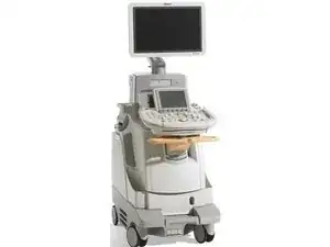
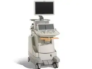
Philips Healthcare produces several different kinds of ultrasound machines including general imaging, cardiovascular, obstetrics and gynecology, and point of care machines. The name “Philips” is generally printed in capital letters at the top of Philips ultrasound machine video monitors. Philips ultrasound monitors are otherwise relatively nondescript but usually include wheels for portability.