
Background and Identification
Magnetic resonance imaging (MRI) is a medical imaging technique used in radiology to form visual images of the anatomy and physiological processes of the body. MRI is a medical application of nuclear magnetic resonance (NMR), which can also be used for imaging in other applications such as NMR spectroscopy. MRI scanners utilize strong magnetic fields, magnetic field gradients, and radio waves to produce images of organs in the body. As opposed to CT (computed tomography scan) and PET (positron emission tomography) scans, MRI does not use X-rays or ionizing radiation.
MRI scans are widely used in hospitals and clinics for medical diagnosis, staging, and follow-up of disease without exposing the body to the radiation of CT scans. MRI scans do include some risks and discomfort as they are longer and louder than CT scans and require the patient to enter a narrow, confining tube. Also, patients with some types of medical implants or non-removable metal inside the body may be unable to undergo an MRI examination safely.
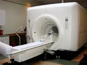
Philips MRI scanners can generally be identified by the name “Philips” printed in black capital letters on the top front of the machine. Philips MRI scanners include a flat surface for the patient to lay on and a large donut-shaped machine with a large hole through the middle that actually takes the images.