Background and Identification
A CT scan or computed tomography scan (previously known as computed axial tomography or CAT scan) is a medical imaging technique that uses computer-processed combinations of multiple X-ray measurements taken from a variety of angles to produce cross-sectional (tomographic) images of a body. A CT scan produces virtual “slices” of a body, allowing users to see inside the body without cutting. The personnel that performs CT scans are called radiographers or radiologic technologists.
Initially, the images generated in CT scans were in the axial anatomical plane, perpendicular to the long axis of the body. Modern CT scanners allow scan data to be reformatted as images in other planes. Digital geometry processing can generate a three-dimensional image of objects inside the body from a series of two-dimensional images taken by rotation around a fixed axis. Such cross-sectional images are widely used for therapy and medical diagnosis.
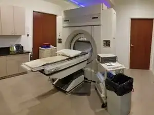
Philips CT scanners can generally be identified by the name “Philips” printed in black capital letters on the top front of the machine. Philips CT scanners include a flat surface for the patient to lay on and a large cuboid-shaped machine with a large hole through the middle that actually takes the images. The patient is passed through the hole in the machine to produce virtual images of the body.

