
Background and Identification
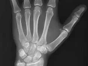
An X-ray or X-radiation is a penetrating form of high-energy electromagnetic radiation. X-ray wavelengths are shorter than those of ultra-violet (UV) rays but longer than those of gamma rays. X-radiation was discovered by Wilhelm Röntgen, a German scientist, in November 1895; in many languages, X-radiation is called Röntgen radiation. Röntgen called it X-radiation to signify an unknown type of radiation. He also discovered that X-rays can identify bone structures, so X-rays are often used for medical imaging. The first medical use of X-rays took place less than one month after Röntgen‘s paper was published on the subject. By 2010, five billion medical imaging examinations had been conducted worldwide using X-rays.
X-rays are used for several medical applications, including projectional radiography, computed tomography (CT scanning), fluoroscopy, and radiotherapy. Fluoroscopy is an imaging technique that uses X-rays to produce real-time moving images of the interior of an object or body. Fluoroscopes are primarily used in medical imaging and allow physicians to see the internal structure of a patient. Fluoroscopes allow doctors to see things like the pumping action of the heart or the motion of swallowing. This is useful for diagnosis and therapy and is used in general radiology, interventional radiology, and image-guided surgery.
GE X-ray machines can generally be identified by the GE emblem, which includes the letters “GE” in cursive font inside of a circle. GE X-ray machines can include a flat surface on which the patient lies and a machine with upper and lower arms that scan the patient. Other X-ray machines are more portable and allow one body part to be scanned.